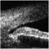
Optical coherence tomography in anterior segment imaging
- PMID: 17355288
- PMCID: PMC2603270
- DOI: 10.1111/j.1600-0420.2007.00876.x
Optical coherence tomography in anterior segment imaging
Abstract
Purpose: To evaluate the ability of optical coherence tomography (OCT), designed primarily to image the posterior segment, to visualize the anterior chamber angle (ACA) in patients with different angle configurations.
Methods: In a prospective observational study, the anterior segments of 26 eyes of 26 patients were imaged using the Zeiss Stratus OCT, model 3000. Imaging of the anterior segment was achieved by adjusting the focusing control on the Stratus OCT. A total of 16 patients had abnormal angle configurations including narrow or closed angles and plateau irides, and 10 had normal angle configurations as determined by prior full ophthalmic examination, including slit-lamp biomicroscopy and gonioscopy.
Results: In all cases, OCT provided high-resolution information regarding iris configuration. The ACA itself was clearly visualized in patients with narrow or closed angles, but not in patients with open angles.
Conclusions: Stratus OCT offers a non-contact, convenient and rapid method of assessing the configuration of the anterior chamber. Despite its limitations, it may be of help during the routine clinical assessment and treatment of patients with glaucoma, particularly when gonioscopy is not possible or difficult to interpret.
Figures


References
-
- Bruno CA, Alward WL. Gonioscopy in primary angle-closure glaucoma. Semin Ophthalmol. 2002;17:59–68. - PubMed
-
- Chan RY, Smith JA, Richardson KT. Anterior segment configuration correlated with Shaffer’s grading of anterior chamber angle. Arch Ophthalmol. 1981;99:104–107. - PubMed
-
- Hoerauf H, Wirbelauer C, Scholz C, Engelhardt R, Koch P, Laqua H, Birngruber R. Slit-lamp-adapted optical coherence tomography of the anterior segment. Graefes Arch Clin Exp Ophthalmol. 2000;238:8–18. - PubMed
-
- Leung CK, Chan WM, Ko CY, Chui SI, Woo J, Tsang MK, Tse RK. Visualization of anterior chamber angle dynamics using optical coherence tomography. Ophthalmology. 2005;112:980–984. - PubMed
-
- Puech M, El Maftouhi A. Exploration du segment anterieur par OCT 3. J Fr Ophtalmol. 2004;27:459–466. - PubMed
Publication types
MeSH terms
Substances
Grants and funding
LinkOut - more resources
Full Text Sources
Other Literature Sources

