. 2007 May 11:1145:117-27.
doi: 10.1016/j.brainres.2007.01.140. Epub 2007 Feb 16.

Midbrain modulation of the cardiac baroreflex involves excitation of lateral parabrachial neurons in the rat

Affiliations

Midbrain modulation of the cardiac baroreflex involves excitation of lateral parabrachial neurons in the rat

Linda F Hayward. Brain Res. .

Abstract

Activation of the dorsal periaqueductal gray (PAG) evokes defense-like behavior including a marked increase in sympathetic drive and resetting of baroreflex function. The goal of this study was to investigate the role of the lateral parabrachial nucleus (LPBN) in mediating dorsal PAG modulation of the arterial baroreflex. Reflex responses were elicited by electrical stimulation of the aortic depressor nerve (ADN) at 5 Hz or 15 Hz in urethane anesthetized rats (n=18). Electrical stimulation of the dorsal PAG at 10 Hz did not alter baseline mean arterial pressure (MAP) but did significantly attenuate baroreflex control of heart rate (HR) evoked by low frequency ADN stimulation. Alternatively, 40 Hz dorsal PAG stimulation increased baseline MAP (43+/-3 mm Hg) and HR (33+/-3 bpm) and attenuated baroreflex control of HR at both ADN stimulation frequencies. Reflex control of MAP was generally unchanged by dorsal PAG stimulation. Bilateral inhibition of neurons in LPBN area (n=6) with muscimol (0.45 nmol per side) reduced dorsal PAG-evoked increases in MAP and HR by 50+/-4% and 95+/-4%, respectively, and significantly reduced, but did not completely eliminate dorsal PAG attenuation of the cardiac baroreflex. Bilateral blockade of glutamate receptors in the LPBN area (n=6) with kynurenic acid (1.8 nmol) had a similar effect on dorsal PAG-evoked increases in MAP, HR and cardiac baroreflex function. Reflex control of MAP was unchanged with either treatment. These findings suggest that the LPBN area is one of several brainstem regions involved in descending modulation of the cardiac baroreflex function during defensive behavior.


Figures

Fig. 1
Fig. 1. The effects of dorsal periaqueductal gray (PAG) stimulation on baroreflex function from one animal
Changes in heart rate (HR), and arterial pressure (AP) during ten seconds of 5 Hz stimulation (horizontal gray bar) of the aortic depressor nerve (ADN; 10V, 2 ms pulse) are shown (A–D). Dashed vertical boxes indicate the two second time windows used for measurement of the effects of ADN stimulation. A. ADN stimulation alone. B. ADN stimulation combined with 10 Hz PAG stimulation (horizontal gray bar). C. ADN stimulation combined with 40 Hz PAG stimulation (horizontal black bar). D. ADN stimulation alone one minute following the offset of 40 Hz PAG stimulation.
Fig. 2
Fig. 2. Averaged effects of 10 versus 40 Hz dorsal PAG stimulation on baroreflex-evoked changes in mean arterial pressure (MAP) and heart rate (HR)
Open bars indicate baroreflex responses evoked by ADN stimulation alone. Hatched bars indicate baroreflex responses evoked by ADN stimulation during 10 Hz PAG stimulation. Black bars indicate responses evoked during 40 Hz PAG stimulation. A. MAP and HR responses during 5 Hz ADN stimulation with or without dorsal PAG stimulation (n=18). B. MAP and HR responses evoked by 15 Hz ADN stimulation with or without dorsal PAG stimulation (n=18). C. MAP and HR responses evoked by 15 Hz ADN stimulation alone (open bar), during 40 Hz PAG (black bar) and one minute following the offset of 40 Hz PAG stimulation (ADN alone, gray bars). Asterisks indicate significantly different (P<0.05) from ADN-alone evoked response.
Fig. 3
Fig. 3. Reconstructed dorsal PAG stimulation sites
Outline of the rostral, middle, and caudal columns of the PAG from Paxinos & Watson’s Rat Brain Stereotaxic Atlas (35). Reconstructed stimulation sites from all animals are organized by experimental group; circles indicate PAG stimulation sites recovered from muscimol microinjection studies; triangles indicate sites from kynurenic acid studies; and rectangles indicate sites from aCSF studies. Note, for illustration purposes all stimulation sites from kynurenic acid experiments are shown on the right side. Numbers refer to midbrain location relative to bregma. Asterisks indicated location of central aqueduct. Dm, dorsomedial column; dl, dorsolateral column; lat, lateral column; vlat, ventrolateral column; Dr, dorsal raphe; Su3, supraoculomotor nucleus; 3mn, oculomotor nucleus.
Fig. 4
Fig. 4. Effects of bilateral inhibition of the lateral parabrachial nucleus (LPBN) on dorsal periaqueductal gray (PAG) modulation of baroreflex function in one animal
Changes in heart rate (HR) and arterial pressure (AP) recorded during 5 Hz stimulation of the aortic depressor nerve (ADN) (horizontal gray bar) from one rat. A. ADN stimulation alone, pre-treatment (left) and ADN stimulation combined with 40 Hz dorsal PAG stimulation (right; horizontal black bar). B. ADN stimulation alone (left) and during 40 Hz dorsal PAG stimulation (right) approximately 5 minutes following bilateral blockade of the LPBN (0.45 nmols muscimol per side).
Fig. 5
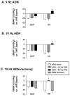
Fig. 5. Averaged effect of dorsal PAG modulation of baroreflex responses before and following lateral parabrachial nucleus (LPBN) microinjection
Open bars indicate baroreflex responses evoked by ADN stimulation alone. Hatched bars indicate baroreflex responses elicited during 40 Hz PAG stimulation before bilateral LPBN blockade. Black bars indicate ADN responses elicited during 40 Hz PAG stimulation after bilateral LPBN blockade. A. MAP and HR responses during 5 Hz ADN stimulation before and after LPBN microinjection (n=6/group). B. Baroreflex responses during 15 Hz ADN stimulation before and after LPBN microinjection (n=6/group). Baroreflex responses evoked alone reflect the average response elicited before and following LPBN microinjection. Left panels, muscimol at 0.45 nmol/side; middle panels, kynurenic acid 1.8 nmol/side; aCSF, 90 nl per side. * indicates significantly different (P<0.05) from ADN-alone evoked response. # indicates significantly different from response evoked by ADN + dorsal PAG stimulation before LPBN microinjection (P<0.05).
Figure 6
Figure 6. Reconstructed lateral parabrachial (LPBN) bilateral microinjection sites
A. Outline of the rostral pons/caudal midbrain from Paxinos & Watson’s (35) and corresponding histological section from one animal illustrating the rostral LPBN viewed with brightfield and fluorescent microscopy. The superior cerebellar peduncle (scp) and boundaries of the LPBN are shown (dashed lines). Arrow indicates the reconstructed center of LPBN microinjection site viewed under fluorescence. B–D. Reconstructed bilateral LPBN microinjection sites (shaded circles) from all muscimol (B, n=6), kynurenic acid (C, n=6), and aCSF (D, n=6), experiments. Asterisks in D illustrate the location of bilateral microinjection sites from one animal made in the ventrolateral pons that did not significantly alter cardiovascular responses evoked from the dorsal PAG and as a result were not included in data analysis. Cross-hatched regions represent location of scp. Numbers to the left of the LPBN images indicate location of the brain section relative to bregma. Aq. indicates location of central aqueduct.

Similar articles

Cited by

References

    1. Bandler R, Shipley MT. Columnar organization in the midbrain periaqueductal gray: Modules for emotional expression? Trends Neurosci. 1994;17:379–389. - PubMed
    1. Bandler R, Carrive P. Integrated defence reaction elicited by excitatory amino acid microinjection in the midbrain periaqueductal grey region of the unrestrained cat. Brain Res. 1988;439:95–106. - PubMed
    1. Bandler R, Prineas S, McCulloch B. Further localization of midbrain neurones mediating the defense reaction in the cat by microinjections of excitatory amino acids. Neurosci Letters. 1985;56:311–316. - PubMed
    1. Cameron AA, Khan IA, Westlund KN, Willis WD. The efferent projections of the periaqueductal gray in the rat: a phaseolus vulgaris-leucoagglutinin study. II Descending Projections. J Comp Neurol. 1995;351:585–601. - PubMed
    1. Carrive PSP, Karli P. Flight induced by microinjection of D-tubocurarine or alpha-bungarotoxin into medial hypothalamus or periaqueductal gray matter: cholinergic or GABAergic mediation? Behav Brain Res. 1986;22 - PubMed

Publication types

MeSH terms