Comparative Study
. 2007 May 4;282(18):13532-41.
doi: 10.1074/jbc.M611295200. Epub 2007 Mar 12.

Coordination of steps in single-nucleotide base excision repair mediated by apurinic/apyrimidinic endonuclease 1 and DNA polymerase beta

Affiliations
Comparative Study

Coordination of steps in single-nucleotide base excision repair mediated by apurinic/apyrimidinic endonuclease 1 and DNA polymerase beta

Yuan Liu et al. J Biol Chem. .

Abstract

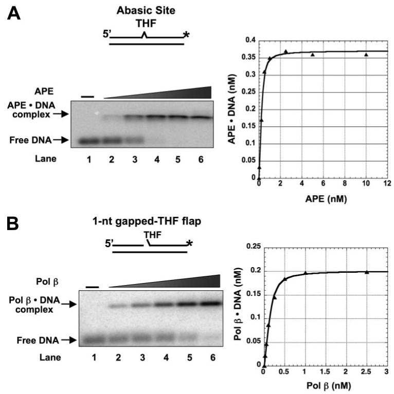
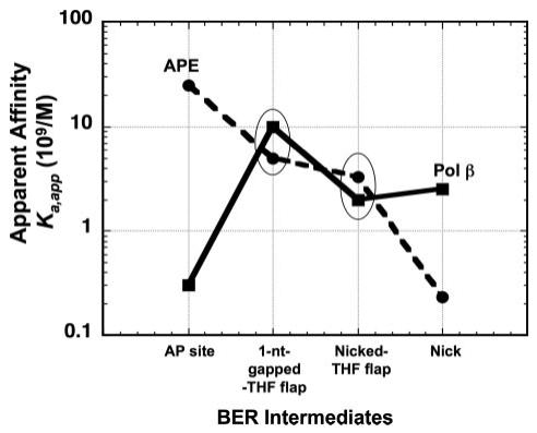
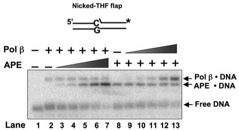
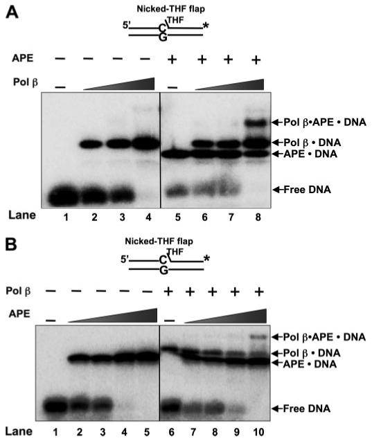
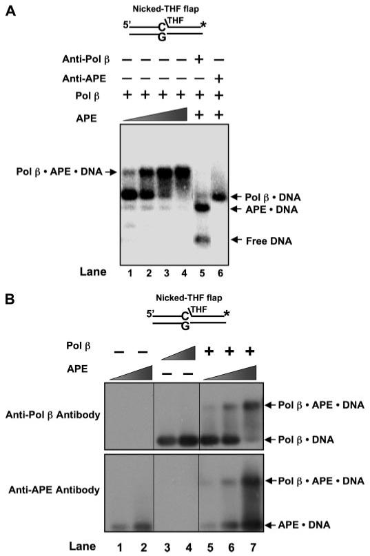
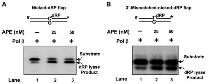
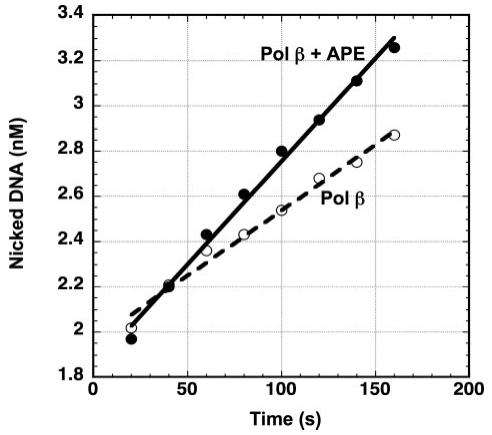
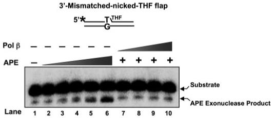
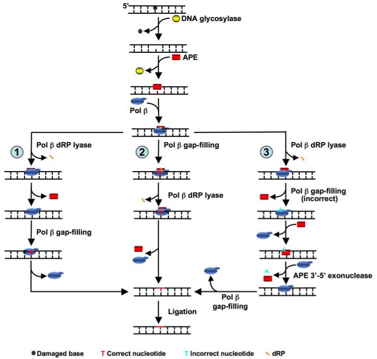
The individual steps in single-nucleotide base excision repair (SN-BER) are coordinated to enable efficient repair without accumulation of cytotoxic DNA intermediates. The DNA transactions and various proteins involved in SN-BER of abasic sites are well known in mammalian systems. Yet, despite a wealth of information on SN-BER, the mechanism of step-by-step coordination is poorly understood. In this study we conducted experiments toward understanding step-by-step coordination during BER by comparing DNA binding specificities of two major human SN-BER enzymes, apurinic/aprymidinic endonuclease 1 (APE) and DNA polymerase beta (Pol beta). It is known that these enzymes do not form a stable complex in solution. For each enzyme, we found that DNA binding specificity appeared sufficient to explain the sequential processing of BER intermediates. In addition, however, we identified at higher enzyme concentrations a ternary complex of APE.Pol beta.DNA that formed specifically at BER intermediates containing a 5'-deoxyribose phosphate group. Formation of this ternary complex was associated with slightly stronger Pol beta gap-filling and much stronger 5'-deoxyribose phosphate lyase activities than was observed with the Pol beta.DNA binary complex. These results indicate that step-by-step coordination in SN-BER can rely on DNA binding specificity inherent in APE and Pol beta, although coordination also may be facilitated by APE.Pol beta.DNA ternary complex formation with appropriate enzyme expression levels or enzyme recruitment to sites of repair.


Figures

FIGURE 1
FIGURE 1. Substrate DNA binding by APE and Pol β
A, a double-stranded DNA with an internal THF residue opposite G in the template strand was employed to measure the binding affinity of APE. The substrate was made by annealing a DNA strand with THF (DS1) to a template strand (T) (see Table 1). The substrate was radiolabeled at the 3′-end of the THF-containing strand (asterisk). Various concentrations of APE (0.05, 0.1, 0.5, 1, and 2.5 nm) were incubated with 0.5 nm substrate at 37 °C in the absence of Mg2+. B, a 1-nt-gapped THF flap substrate was used to examine Pol β substrate binding. The substrate was constructed by annealing an upstream primer (Ugap), a downstream oligomer (DTHF-flap) to the template strand (T). The substrate was radiolabeled at the 3′-end of the downstream oligonucleotide. Pol β (0.05, 0.1, 0.2, 0.25, and 0.5 nm) was incubated with 0.5 nm substrate at 37 °C for 8 min. After incubation, the protein-DNA complex was separated from free DNA by agarose-acrylamide gel electrophoresis as described under “Experimental Procedures.” The binding curves of the enzymes are shown in the right panels. The DNA substrates are schematically illustrated above each gel photograph.
FIGURE 2
FIGURE 2. Binding affinity of APE and Pol β to various SN-BER intermediates
The apparent binding affinities (Ka,app; 1/Kd,app) of APE and Pol β to a double-strand DNA with an abasic site, a 1-nt-gapped THF flap, a nicked-THF flap, and a nicked DNA (Table 2) were plotted as a function of the sequential BER intermediate steps during simple SN-BER. The oval circles highlight the intermediates to which Pol β and APE exhibit similar apparent binding affinities.
FIGURE 3
FIGURE 3. APE and Pol β compete for a nicked-THF flap BER intermediate
Increasing concentrations of APE (lanes 3-7) and Pol β (lanes 9-13) were incubated with 0.25 nm Pol β or 0.25 nm APE and with 0.5 nm substrate DNA. The binding assay was performed with the conditions described under “Experimental Procedures.” Lanes 3-7 include 0.05, 0.1, 0.25, 0.5, and 1 nm APE along with 0.25 nm Pol β, whereas lanes 9-13 include the same concentrations of Pol β along with 0.25 nm APE. Lane 2 was the mixture that contained only Pol β and substrate DNA, whereas lane 8 was the mixture that contained APE and substrate DNA. Lane 1 is substrate DNA alone. The substrate is schematically illustrated above the gel.
FIGURE 4
FIGURE 4. Formation of APE·Pol β·DNA ternary complex
A, increasing concentrations of Pol β were incubated with 5 nm nicked-THF flap substrate DNA in the absence (lanes 2-4) and presence of 1 nm APE (lanes 6-8). Lane 1 represents the binding mixture without enzyme. Lanes 2-4 correspond to mixtures containing increasing concentrations of Pol β (0.5, 1, and 2.5 nm, respectively) and DNA. Lanes 6-8 indicate mixtures containing the same concentrations of Pol β as lanes 2-4 along with 1 nm APE. Lane 5 indicates a binding mixture with APE and the DNA substrate. B, increasing concentrations of APE (0.5, 1, 2.5, and 5 nm, respectively) were incubated with 5 nm nicked-THF flap substrate DNA in the absence (lanes 2-5) and presence of 1 nm Pol β (lanes 7-10). Lane 1 represents the reaction mixture without enzymes, whereas lane 6 corresponds to the mixture containing Pol β and DNA substrate. The enzymes were incubated with the DNA substrates with the conditions described under “Experimental Procedures,” and the protein-DNA complexes were analyzed. The substrate DNA is schematically illustrated above the gel.
FIGURE 5
FIGURE 5. Identification of components of the APE·Pol β·DNA ternary complex
The co-existence of APE and Pol β in the ternary complex was probed by using specific polyclonal antibody against either APE or Pol β and immunoblotting. A, each antibody was incubated with the enzymes and 5 nm nicked-THF flap substrate with the conditions described under “Experimental Procedures.” Lanes 1-4 represent mixtures containing various concentrations of APE (1, 2.5, 10, 25 nm, respectively), 1 nm Pol β, and DNA. Lane 5 corresponds to the mixture containing Pol β, APE, the specific antibody against Pol β (∼0.3 μg/μl), and the DNA substrate, whereas lane 6 represents the mixture with APE, Pol β, specific antibody against APE (∼0.3 μg/μl), and the DNA substrate. The substrate is schematically illustrated above the gel. B, the protein-DNA complexes were initially separated from free DNA by electrophoresis and transferred to PVDF membrane by the vacuum-dry procedure, and the membrane was immunoblotted as described under “Experimental Procedures.” The upper panel illustrates immunoblotting results with anti-Pol β antibody, and the lower panel represents the blot with anti-APE antibody. Lanes 1 and 2 are reactions with 10 and 20 nm APE, respectively, whereas lanes 3 and 4 represent mixtures with 10 and 20 nm Pol β, respectively. Lanes 5-7 represent mixtures of 20 nm Pol β and 10, 20, or 30 nm APE, respectively. The substrate is schematically illustrated above the gel.
FIGURE 6
FIGURE 6. APE·Pol β·DNA ternary complex stimulates Pol β lyase activity
Pol β (2 or 5 nm) was incubated with 10 nm 3′-matched (A) or mismatched (B) nicked-dRP flap substrate in the absence (lane 1) and presence of 25 and 50 nm APE (lanes 2 and 3) at 30 °C for 10 min. The reaction mixtures were incubated and quenched as described under “Experimental Procedures.” The product and substrate were then separated by denaturing polyacrylamide gel electrophoresis and measured as described under “Experimental Procedures.” The nickeddeoxyuridine flap substrate was constructed by annealing the 3′-end radiolabeled (asterisk) downstream oligonucleotide and the upstream primer to the template strand. The dRP lyase substrates were prepared by treating with uracil DNA glycosylase just before performing the lyase assay. The substrate is schematically illustrated above the gel.
FIGURE 7
FIGURE 7. Stimulation of Pol β 1-nt gap-filling activity by APE·Pol β·DNA ternary complex
The rate of Pol β 1-nt gap-filling activity was measured with the conditions of APE·Pol β·DNA complex formation as described under “Experimental Procedures.” The reaction was performed in the presence of 1 nm Pol β or 1 nm Pol β and 25 nm APE, 5 mm MgCl2,50 μm dCTP, and 10 nm DNA substrate. Ten-μl aliquots were collected at timed intervals (0.5-5 min) and quenched with 250 mm EDTA. The data were fitted to a linear equation, and the rate was obtained from the slope.
FIGURE 8
FIGURE 8. APE·Pol β·DNA ternary complex does not affect the APE 3′-5′ exonuclease activity
Mismatched-nicked-THF flap substrate (10 nm) was incubated either with increasing concentrations of APE (5, 25, 50 nm) (lanes 2-6, respectively) or with 2.5 nm APE and increasing concentrations of Pol β (5-50 nm) (lanes 7-10, respectively) at 37 °C for 15 min as described under “Experimental Procedures.” Lane 1 was the reaction with only substrate DNA. The product and substrate were separated by denaturing polyacrylamide gel electrophoresis as described under “Experimental Procedures.” The substrate was made by annealing the 5′-end radiolabeled (asterisk) upstream primer and the downstream oligomer to the template. The substrate is schematically illustrated above the gel.
FIGURE 9
FIGURE 9. Mechanism of SN-BER repair as function of substrate binding specificity and protein-protein interactions
DNA glycosylase removes a damaged base and leaves an abasic site. APE binds and makes a 5′-incision adjacent to the AP site leaving a 1-nt gap with a 5′-dRP residue. With a high local enzyme concentration, APE and Pol β can form a protein-DNA ternary complex on this intermediate that stimulates Pol β activities (1-nt gap-filling and dRP lyase). Three subpathway choices may occur at this point; in one scenario, as indicated in subpathway 1, Pol β dRP lyase activity stimulated by APE·Pol β·DNA ternary complex removes the dRP group, generating a 1-nt-gapped DNA intermediate. The lower binding affinity of APE for this intermediate may promote dissociation of APE, whereas Pol β effectively binds to this intermediate and fills the gap producing the nicked DNA substrate necessary for ligation. In another scenario, the protein-DNA ternary complex with Pol β performs 1-nt gap-filling DNA synthesis before it removes the dRP group, thereby creating a nicked-dRP flap intermediate (subpathway 2). Under high local enzyme concentrations, the ternary complex may dissociate before the next step in the pathway. However, when a dRP-containing intermediate is present, APE and Pol β can rapidly reform the ternary complex (Table 2). These dissociation and re-association steps are not explicitly illustrated to simplify the overall scheme. The Pol β dRP lyase then removes the sugar-phosphate residue, creating nicked DNA for ligation. In subpathway 3, Pol β removes the dRP group before misinserting a nucleotide, generating a nicked intermediate with a 3′-mismatch. APE 3′-exonuclease activity can then remove the 3′-misinserted nucleotide. This results in 1-nt-gapped DNA that Pol β will efficiently insert the correct nucleotide, creating a nicked DNA that can then be ligated.

References

    1. Lindahl T, Ljungquist S. Basic Life Sci. 1975;5:31–38. - PubMed
    1. Ames BN. Free Radic. Res. Commun. 1989;7:121–128. - PubMed
    1. Ames BN, Gold LS. Mutat. Res. 1991;250:3–16. - PubMed
    1. Lutz WK. Mutat. Res. 1990;238:287–295. - PubMed
    1. Lindahl T. Nature. 1993;362:709–715. - PubMed

Publication types

MeSH terms