

Characterization of human corneal stem cells by synchrotron infrared micro-spectroscopy
- PMID: 17356510
- PMCID: PMC2633470
Characterization of human corneal stem cells by synchrotron infrared micro-spectroscopy
Abstract
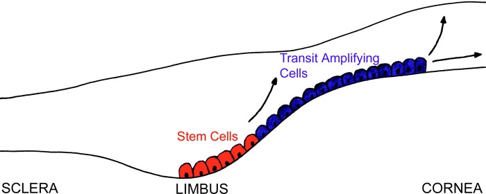
Purpose: The purpose of this study was to use high resolution synchrotron radiation-based Fourier Transform Infrared (FTIR) micro-spectroscopy coupled with multivariate analysis to investigate the characteristics of adult stem cell (SC) and transit amplifying (TA) cell populations of the human corneal epithelium.
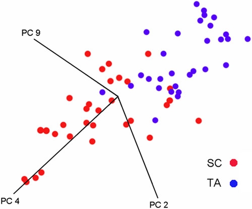
Methods: Spectra of individual SC and TA cells in situ from cryosections of human cornea were collected using a synchrotron micro-spectroscopy facility at Daresbury laboratory, UK. Multivariate analysis and Mann Whitney U tests were used to analyse the spectral data from the SC and TA cell populations.
Results: There were marked differences between the median spectra of the two cell populations. This correlated with their level of differentiation and functional specialization. Multivariate (principal component) analysis revealed that the cell populations could be segregated into distinct clusters, with only slight overlap between the two cell types. Significant (p<0.05) spectral differences were found in the spectral regions associated with nucleic acid, protein and lipids.
Conclusions: Synchrotron FTIR micro-spectroscopy together with principal component analysis is able to discriminate between SC and TA cell populations. Our results also suggest a small sub-population of corneal epithelial cells in the SC niche have TA cell-like characteristics. Many of the spectral differences between the SC and TA cell populations relate to differences in nucleic acid conformation.
Figures

References
-
- Hall PA, Watt FM. Stem cells: the generation and maintenance of cellular diversity. Development. 1989;106:619–33. - PubMed
-
- Davanger M, Evensen A. Role of the pericorneal papillary structure in renewal of corneal epithelium. Nature. 1971;229:560–1. - PubMed
-
- Cotsarelis G, Cheng SZ, Dong G, Sun TT, Lavker RM. Existence of slow-cycling limbal epithelial basal cells that can be preferentially stimulated to proliferate: implications on epithelial stem cells. Cell. 1989;57:201–9. - PubMed
-
- Lavker RM, Tseng SC, Sun TT. Corneal epithelial stem cells at the limbus: looking at some old problems from a new angle. Exp Eye Res. 2004;78:433–46. - PubMed
Publication types
MeSH terms
Substances
LinkOut - more resources
Full Text Sources
Medical