. 2007 Feb 26:13:258-72.

Retinal pathology and skin barrier defect in mice carrying a Stargardt disease-3 mutation in elongase of very long chain fatty acids-4

Affiliations

Retinal pathology and skin barrier defect in mice carrying a Stargardt disease-3 mutation in elongase of very long chain fatty acids-4

Anne McMahon et al. Mol Vis. .

Abstract

Purpose: Autosomal dominant Stargardt disease-3 (STGD3) is caused by mutations in elongase of very long chain fatty acids-4 (ELOVL4). The goal of this study was to generate and characterize heterozygous and homozygous knockin-mice that carry a human STGD3 pathogenic mutation in the mouse Elovl4 gene.

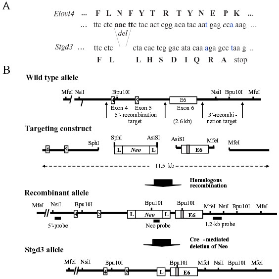
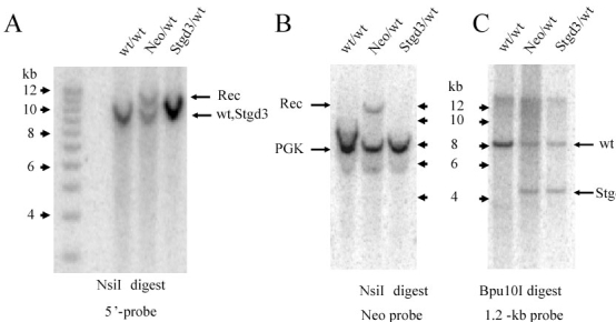
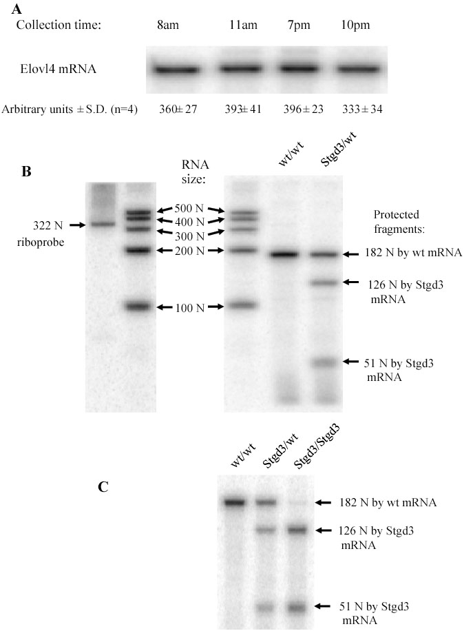
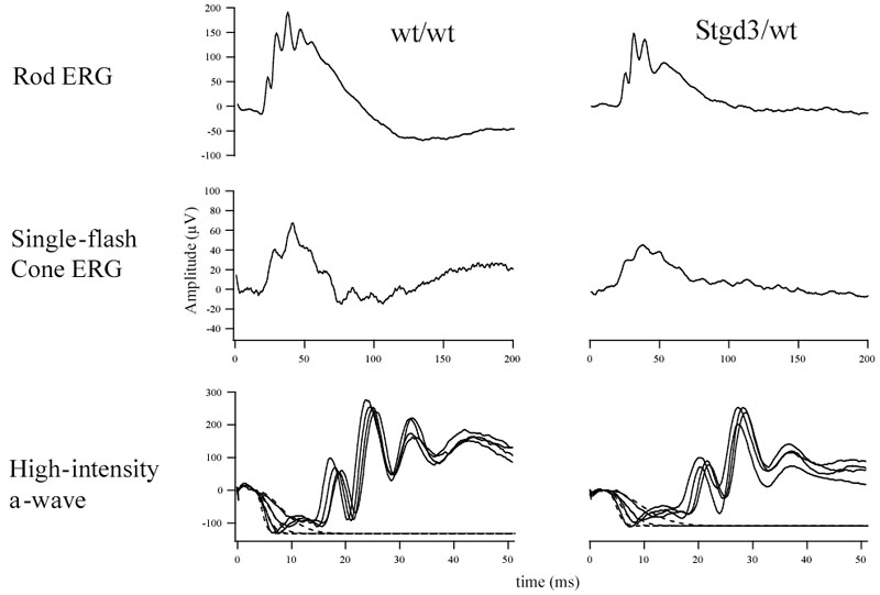
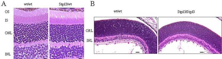
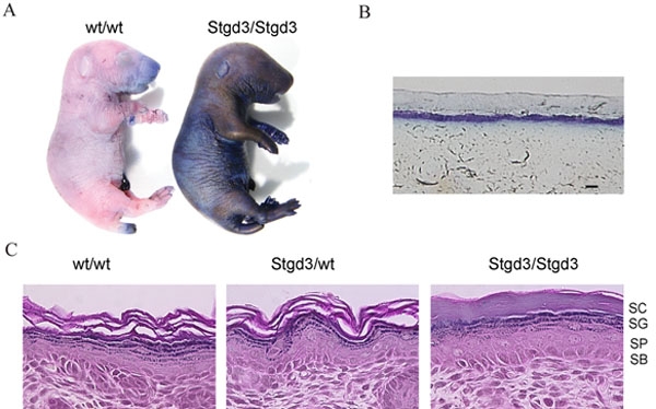
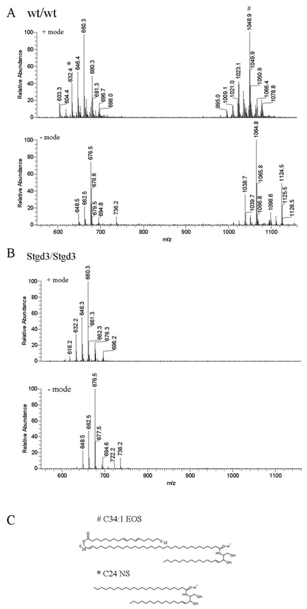
Methods: Recombinant Stgd3-knockin mice were generated using a DNA construct which introduced a pathogenic five-base pair deletion and two point mutations in exon 6 of the Elovl4 gene. Stgd3-mouse genotypes were confirmed by Southern blot analysis and expression of wild-type (wt) and mutated Elovl4 mRNAs assayed by nuclease protection assay. The retinal phenotype of heterozygous Stgd3 mice was characterized by morphological studies, elecroretinographic (ERG) analysis and assay of lipofuscin accumulation. Homozygous Stgd3 mice were examined for both retinal and gross morphology. They were also analyzed for skin morphology and skin barrier function, and for epidermal lipid content using high performance liquid chromatography (HPLC) combined with mass spectrometry (MS).

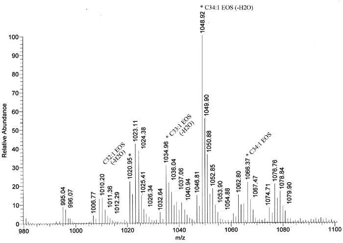
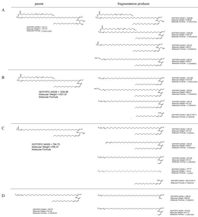
Results: The Stgd3 allele codes for a truncated mouse Elovl4 protein, which also contains the same aberrant 8-amino acid C-terminus encoded by the human pathogenic STGD3 allele. Heterozygous Stgd3 mice expressed equal amounts of both wt and mutant Elovl4 mRNAs in the retina, showed no significant changes in retinal morphology, but did show accumulation of lipofuscin and reduced visual function. Homozygous Stgd3 mice were born with an expected Mendelian frequency, without any initial gross anatomical or behavioral abnormalities. By 6-12 h postpartum, they became dehydrated and died. A skin permeability assay detected a defect in epidermal barrier function. Homozygous mutant epidermis expressed a normal content of mutated Elovl4 mRNA and contained all four epidermal cellular layers. HPLC/MS analysis of epidermal lipids revealed the presence of all barrier lipids with the exception of the complete absence of acylceramides, the critical lipids for barrier function of the skin.

Conclusions: The generated Stgd3-knockin mice are a genetic model of human STGD3 and reproduce features of the human disease: accumulation of lipofuscin and reduced visual functions. Homozygous Stgd3 mice showed a complete absence of acylceramides from the epidermis. Their absence suggests a role for Elovl4 in acylceramide synthesis, and in particular, a role in the synthesis of the unique very long chain C30-C40 fatty acids present in skin acylceramides.


Figures

Figure 1
Figure 1
Gene-targeting strategy for generation of Stgd3-gene knockin mice. A: Partial nucleotide sequence from exon 6, and the encoded amino acids (capital letters), of the wt (Elovl4) and mutant (Stgd3) alleles. Shown are the mutations introduced into the Elovl4 sequence prior to assembly of the targeting construct. Five nucleotide base pairs, corresponding to those absent in Stargardt-3 patients, were deleted (del). Two point mutations (the altered and substituted nucleotides are shown in blue) were introduced in the sequence downstream of the deletion to generate a new C-terminal sequence that is identical to that found in STGD3 patients. B: Schematic maps of a portion of the Elovl4 allele encompassing exons 4, 5, and 6, the targeting construct, recombinant allele and the final Stgd3 allele. Restriction sites used for cloning and for Southern blot verification of recombination and deletion events, as well as the location of the Southern probes are included. Mutations in exon 6 are shown schematically as a shaded box, within which the new Bpu10I restriction site introduced during sequence manipulation is shown. Homologous recombination of the 11.5-kb targeting construct generates a recombinant allele containing exon 6 with the Stgd3 mutation, a neomycin selection cassette (Neo) flanked by lox P sites (L), and 4.2 kb and 3.1kb of 5'- and 3'-targeting sequence, respectively. Breeding of recombinant Neo/wt mice with Cre-transgenic mice leads to Cre-mediated deletion of the Neo cassette and generation of the mice carrying the final Stgd3 allele.
Figure 2
Figure 2
Southern blot verification of Stgd3 mouse genotype. A: Genomic DNA was digested with NsiI and probed with a 5'-probe located upstream of the recombinant targeted site (see Figure 1). A signal from the wt allele as well as an extra band (Rec), derived from the recombinant allele, were detected in the Neo/wt DNA digests. The size of the Rec band was consistent with the 11.8-kb size predicted for the homologous recombination event, confirming the presence of recombination in these mice. In the Stgd3/wt DNA digests, the size of the recombined band was reduced due to loss of the Neo cassette with the result that the wt and Stgd3 allele-derived signals were not resolved. B: Hybridization with the Neo probe detected the presence of the Neo signal in the Neo/wt but not in Stgd3/wt DNA digests, confirming recombination of the targeting sequence and Cre-mediated excision of the Neo cassette, respectively. The Neo probe contained a mouse phosphoglycerate kinase (PGK) promoter and thus also detected the endogenous PGK-gene sequence in all mouse DNA samples. C: Introduction of the STGD3 mutation into Elovl4 exon 6 generated a new Bpu10I restriction site in this exon. The presence of the STGD3-mutation in the DNA of Neo/wt and Stgd3/wt mice was confirmed by the detection of a new 4.3-kb Bpu10I restriction fragment that was absent from wt/wt DNA.
Figure 3
Figure 3
Expression of Elovl4 and Stgd3 mRNAs in mouse eyecups. S1-nuclease protection assay, using a 322-nucleotide (N) riboprobe, detected a 182-N fragment protected by Elovl4 (wt) mRNA and two fragments (126 Ns and 51 Ns) protected by Stgd3 (mutant) mRNA. A: Elovl4 mRNA levels in wt/wt retinas remained constant through the day/night cycle. B: Stgd3/wt retinas from 1-month-old mice expressed equivalent amounts of both wt and Stgd3 mRNAs (ratio 1.02±0.06; mean±SD, n=5). The level of Elovl4 mRNA in these mice was a half (51%±6; mean±SD, n=4) of that detected in the wt/wt littermates. RNA size standards in a range of 100 N-500 N are shown. C: The epidermal level of mutated Elovl4 mRNA (Stgd3 mRNA) in neonatal Stgd3/Stgd3 mice is comparable to the Elovl4 mRNA levels in wt/wt and Stgd3/wt littermates.
Figure 4
Figure 4
Electroretinographic analysis of heterozygous Stgd3 mice. Representative rod and cone electroretinograms, and a-wave responses are presented for 8-month-old Stgd3/wt mice and their wt/wt littermates. Electroretinographic parameters obtained from all mice (n=6 per group) examined for the study are summarized in Table 2.
Figure 5
Figure 5
Light micrographs of hematoxylin and eosin stained sections of Stgd3 mouse retinas. A: Retinal sections from 8-month-old heterozygous Stgd3 mice and their wt littermates show similar thickness of photoreceptor outer segments (OS), inner segment (IS), outer nuclear (ONL) and inner nuclear (INL) layers. No significant changes in numbers and morphology of photoreceptors are evident. Scale bar represents 10 μm. B: Retinal sections from neonatal homozygous Stgd3 mice and their wt littermates show similar retinal histology, with distinct INL and larger outer retinal layers (ORL). The ORL contains the photoreceptor cell bodies which at this stage of development have not elaborated outer segments. Scale bar represents 100 μm.
Figure 6
Figure 6
Defective skin barrier in neonatal homozygous Stgd3 mice. A: Skin permeability assay. Euthanized neonatal pups were immersed in a toluidine blue solution. The skin of homozygous Stgd3 mice, in contrast to their wt littermates, stained blue indicating a defect in skin barrier function. B: Light microscopy examination of a cross section of the dye-stained skin of the homozygous Stgd3 neonate shows penetration of the dye through the outer skin layers. C: Hematoxylin and eosin staining of homozygous Stgd3 skin shows the presence of all four epidermal layers: stratum corneum (SC), stratum granulosum (SG), stratum spinosum (SP) and stratum basale (SB). Note the more compact structure of stratum corneum in the mutant homozygous skin compared to the wt/wt and Stgd3/wt skin. Scale bar represents 100 μm in B.
Figure 7
Figure 7
Epidermal ceramides in neonatal homozygous Stgd3 mice. Epidermal lipids were extracted and analyzed by high performance liquid chromatography/mass spectrometry (HPLC/MS). Full scan MS analysis of an HPLC ceramide fraction detected non-acylceramides (600-700 m/z) in both wt/wt (A) and Stgd3/Stgd3 epidermal extracts (B), but acylceramides (1000-1100 m/z) only in the wt/wt extract. Among non-acylceramides, we identified C24-NS ceramide based on a comparison with a commercial C24-NS standard (isotopic mass=649.63). M/z values for both were identical; 648.5 m/z in negative ion mode due to proton loss, and 632.4 m/z in positive ion mode due to proton gain and water loss. Also detected in both extracts were its CH2-homologs: C25-NS ceramide (662.5 and 646.4 m/z in negative and positive mode, respectively) and C26-NS ceramide (676.5 and 660.3 m/z, respectively). Among acylceramides in the wt/wt extract, we identified C34:1-EOS ceramide (isotopic mass 1066.00) based on an m/z value of 1064.8 in negative mode due to proton loss, and 1048.9 m/z in positive mode due to proton gain and water loss. Identity was further confirmed by fragmentation analysis (see Figure 9). Also present were CH2-homologs of C34-EOS containing varying degrees of fatty acid saturation (see Figure 8). C shows the structures of an acylceramide with a C34-fatty acid (C34:1-EOS) and a non-acylceramide with a C24 fatty acid (C-24-NS).
Figure 8
Figure 8
Acylceramides of wild-type mouse epidermis. Extended print-out of the acylceramides detected using positive ion mode MS analysis (Figure 7). It shows the 1066.37 m/z peak identified as C34:1-EOS by fragmentation analysis (see Figure 9) and the 1048.92 m/z peak of its dehydrated derivative. Signals 1020.95, and 1034.96 m/z correspond to C32:1-EOS, and C33:1-EOS, respectively. These are CH2-homologs of C34-EOS. Other peaks can be assigned as EOS ceramides containing varied degrees of fatty acid saturation or as minor acylceramide species (EOH and EOP) that contain an additional hydroxy group.
Figure 9
Figure 9
Fragmentation analysis of wild-type epidermal ceramide. The MS positive mode 1066.4 m/z peak (see Figure 7A) was fragmented by collision-induced dissociation and resulted in generation of 1048.1, 1030.6, 744.2, and 769.0 m/z daughter ions. Chemical structures for compounds of these molecular masses are provided in panel A: Further fragmentation of the 1030 m/z ion produced 1012.9, 768.6, 750.7, and 263.9 m/z signals (deduced compound formulas are shown in panel B). Transformation of the 768.6 m/z ion yielded 750.5 + 708.7 + 570.3 + 444.3 + 263.9 m/z ions (C), and fragmentation of the 750.5 m/z ion produced 571.1 and 506.2 m/z ions (D). The observed daughter ions are consistent with fragment compounds expected from C34:1-EOS ceramide which contains sphingosine linked to an ω-hydroxy C34 monounsaturated fatty acid that is esterified with linoleic acid.

References

    1. Zhang K, Kniazeva M, Han M, Li W, Yu Z, Yang Z, Li Y, Metzker ML, Allikmets R, Zack DJ, Kakuk LE, Lagali PS, Wong PW, MacDonald IM, Sieving PA, Figueroa DJ, Austin CP, Gould RJ, Ayyagari R, Petrukhin K. A 5-bp deletion in ELOVL4 is associated with two related forms of autosomal dominant macular dystrophy. Nat Genet. 2001;27:89–93. - PubMed
    1. Edwards AO, Donoso LA, Ritter R., 3rd A novel gene for autosomal dominant Stargardt-like macular dystrophy with homology to the SUR4 protein family. Invest Ophthalmol Vis Sci. 2001;42:2652–63. - PubMed
    1. Bernstein PS, Tammur J, Singh N, Hutchinson A, Dixon M, Pappas CM, Zabriskie NA, Zhang K, Petrukhin K, Leppert M, Allikmets R. Diverse macular dystrophy phenotype caused by a novel complex mutation in the ELOVL4 gene. Invest Ophthalmol Vis Sci. 2001;42:3331–6. - PubMed
    1. Maugeri A, Meire F, Hoyng CB, Vink C, Van Regemorter N, Karan G, Yang Z, Cremers FP, Zhang K. A novel mutation in the ELOVL4 gene causes autosomal dominant Stargardt-like macular dystrophy. Invest Ophthalmol Vis Sci. 2004;45:4263–7. - PubMed
    1. Stone EM, Nichols BE, Kimura AE, Weingeist TA, Drack A, Sheffield VC. Clinical features of a Stargardt-like dominant progressive macular dystrophy with genetic linkage to chromosome 6q. Arch Ophthalmol. 1994;112:765–72. - PubMed

Publication types

MeSH terms

Associated data

LinkOut - more resources