Comparative Study
. 2007 Apr;80(4):616-32.
doi: 10.1086/512735. Epub 2007 Feb 14.

Disruption of ROBO2 is associated with urinary tract anomalies and confers risk of vesicoureteral reflux

Affiliations
Comparative Study

Disruption of ROBO2 is associated with urinary tract anomalies and confers risk of vesicoureteral reflux

Weining Lu et al. Am J Hum Genet. 2007 Apr.

Abstract

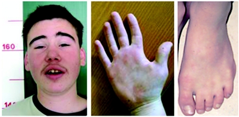
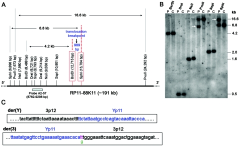
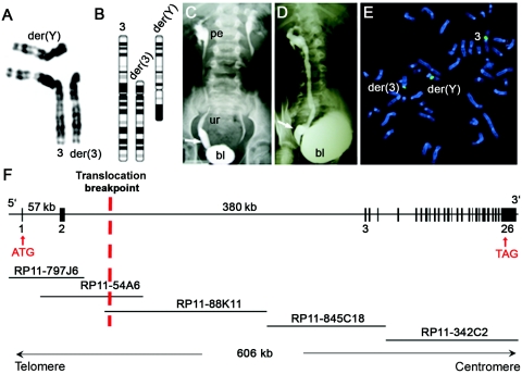
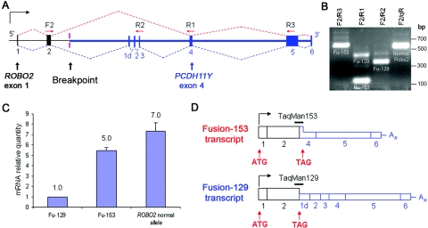
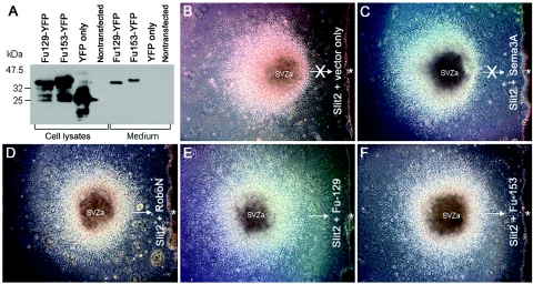
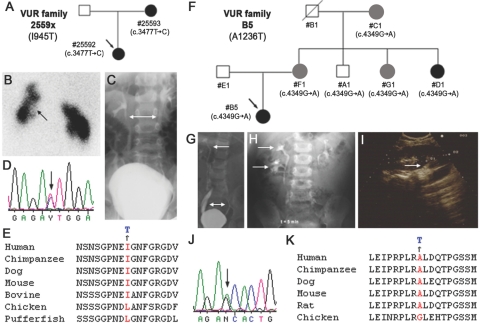
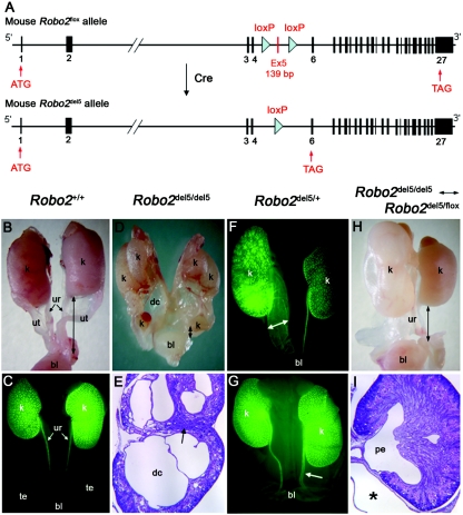
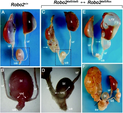
Congenital anomalies of the kidney and urinary tract (CAKUT) include vesicoureteral reflux (VUR). VUR is a complex, genetically heterogeneous developmental disorder characterized by the retrograde flow of urine from the bladder into the ureter and is associated with reflux nephropathy, the cause of 15% of end-stage renal disease in children and young adults. We investigated a man with a de novo translocation, 46,X,t(Y;3)(p11;p12)dn, who exhibits multiple congenital abnormalities, including severe bilateral VUR with ureterovesical junction defects. This translocation disrupts ROBO2, which encodes a transmembrane receptor for SLIT ligand, and produces dominant-negative ROBO2 proteins that abrogate SLIT-ROBO signaling in vitro. In addition, we identified two novel ROBO2 intracellular missense variants that segregate with CAKUT and VUR in two unrelated families. Adult heterozygous and mosaic mutant mice with reduced Robo2 gene dosage also exhibit striking CAKUT-VUR phenotypes. Collectively, these results implicate the SLIT-ROBO signaling pathway in the pathogenesis of a subset of human VUR.


Figures

Figure  A1.
Figure A1.
Facial and limb abnormalities of the DGAP107 proband. Note the low-set, dysplastic ears and subtle membranous syndactyly and clinodactyly. Blepharophimosis is also present.
Figure  B1.
Figure B1.
ROBO2 disrupted in DGAP107, with the breakpoint lying within intron 2. A, Restriction map surrounding the 3p12 breakpoint. The base-pair position of BAC RP11-88K11 (AC131005, within intron 2 of ROBO2 [see BAC contig in fig. 1F]) was used to calculate the distance between restriction enzyme sites. RP11-88K11 overlaps with BAC clone RP11-54A6 used in FISH and also contains the breakpoint, which is between boxed BsrDI and SphI sites, on the basis of the aberrant bands detected by Southern blot analysis. B, Southern blot analysis of DGAP107 (P) and unaffected control (C) genomic DNA, with use of the designated restriction enzymes and the probe A2-57 shown in panel A. Aberrant bands (white arrows) are present only in DGAP107 DNA digested with SphI and PvuII. C, Breakpoint cloning showing the sequence of the junction fragment from der(3) with a 1-bp deletion (g [green]) and a 2-bp insertion (tt [pink]). There is no gain or loss of nucleotides at the der(Y) breakpoint.
Figure  C1.
Figure C1.
Expression of ROBO2 and PCDH11Y in human tissues. RT-PCR amplified 2.8-kb ROBO2 cDNAs with the use of primers ROBO2-F1 and ROBO2-R1. The 3-kb ROBO2 cDNA product (upper band of doublet) contains an alternatively spliced exon 24B. RT-PCR amplification of 404-bp and 620-bp PCDH11Y cDNAs used primers PCDH11Y-F1 and PCDH11Y-R1. The intensity of the fragments indicates the approximate expression level of ROBO2 and PCDH11Y in these tissues. Notably, there is no expression of PCDH11Y in the fetal kidney. β-actin was used as a cDNA loading control.
Figure  F1.
Figure F1.
Generation of F2 Robo2del5/del5Robo2del5/flox mosaics
Figure  1.
Figure 1.
ROBO2 disrupted in DGAP107. Partial karyogram (A) and idiogram (B) for 46,X,t(Y;3)(p11;p12)dn is shown. VCUG of DGAP107 shows anterior-posterior (C) and lateral (D) views of bilateral grade IV VUR and megaureter at the right UVJ (arrows). bl = Bladder; pe = renal pelvis; ur = ureter. E, FISH analysis showing BAC RP11-54A6 (green), which hybridizes to normal chromosome 3, der(3), and der(Y) and crosses the 3p12 breakpoint. F, Intron-exon structure of ROBO2, with select exons numbered and the relevant BAC contig. The location of the 3p12 translocation breakpoint is indicated by a red dotted vertical line.
Figure  2.
Figure 2.
The t(Y;3) translocation in DGAP107, which generates novel ROBO2 fusion transcripts. A, ROBO2 and PCDH11Y intron-exon structure surrounding the der(Y) breakpoint. The forward primer F2 in ROBO2 exon 2 (black bar) was used in RT-PCR with three reverse primers—R2, R1, and R3—in PCDH11Y exons 3, 4, and 5, respectively (blue bars). Dotted lines indicate the observed splicing patterns of the two fusion transcripts. The red splicing pattern generates Fu-153, which encodes 153 aa, and the blue pattern generates Fu-129, which encodes 129 aa. B, RT-PCR fusion transcript amplification. Lane 1, F2/R3 primers amplify Fu-153 (641 bp) and Fu-129 transcripts; only the shorter Fu-153 amplicon is shown. Lane 2, F2/R1 primers amplify transcripts for both Fu-129 (456 bp) and Fu-153 (122 bp). Lane 3, F2/R2 primers amplify only Fu-129 transcripts (347 bp). Lane 4, F2/qR primers amplify only transcripts from the wild-type nontranslocated ROBO2 allele (606 bp). qR primer is located in exon 7 of ROBO2. C, Real-time RT-PCR quantitation of ROBO2 fusion transcripts Fu-129 and Fu-153 (detected by TaqMan probes shown in panel D) and of ROBO2 nontranslocated allele transcripts (detected by TaqMan probe across ROBO2 exons 2 and 3) in DGAP107 lymphoblast RNA. D, Exon structure of Fu-153 and Fu-129. Horizontal bars indicate TaqMan probes used to quantify fusion transcripts. Black boxes indicate ROBO2 exons; blue boxes, PCDH11Y exons; full-height boxes, coding exons; and half-height boxes, noncoding exons.
Figure  3.
Figure 3.
ROBO2 fusion proteins inhibiting SLIT chemorepulsion. A, YFP-tagged ROBO2 fusion proteins (Fu129-YFP [40 kDa] and Fu153-YFP [42 kDa]) detected by an anti-YFP antibody, expressed in HEK cell lysates, and secreted into the medium. In the presence of aggregated cells transfected with Slit2 plus empty vector (B) or Slit2 plus Sema3A (Semaphorin 3A, with no effect on Slit2 repulsive activity) (C), cells migrate out of SVZa explants and away from the Slit2-expressing cell aggregate (asterisk). In the presence of aggregated cells transfected with Slit2 plus RoboN (the Robo extracellular domain, which inhibits Slit repulsive activity), cells migrate out of SVZa explants symmetrically in all directions (D) including toward (arrow) the Slit2 and RoboN-expressing cell aggregate (asterisk). Fu-129 and Fu-153 also effectively block Slit2 repulsive activity (E and F), allowing symmetrical neuronal migration out of SVZa explants and toward (arrows) Slit2 and Fu-129 or Slit2– and Fu-153–expressing cell aggregates (asterisks).
Figure  4.
Figure 4.
ROBO2 missense mutations in familial CAKUT and VUR. A, Family 2559x with CAKUT-VUR and exon 19 (c.3477T→C) mutation. Arrow indicates the proband. Blackened and gray symbols indicate patients with CAKUT-VUR and family members with urinary tract symptoms and radiological evidence of CAKUT. Nucleotide changes are shown under each individual. B, 99mTc-dimercaptosuccinic acid renogram of proband 25592 showing bilateral renal parenchymal defects (arrow). C, VCUG of proband 25592 showing bilateral reflux (bidirectional arrow). Chromatograms show T→C change (arrow) in exon 19 of family 2559x (D) and amino acid conservation across species (E). F, Family B5 with CAKUT-VUR and exon 23 (c.4349G→A) mutation. G, VCUG showing bilateral VUR (bidirectional arrow) and right duplex kidney (arrow) in proband B5. H, IVP detecting right duplex kidney (arrows) in proband B5. I, US showing suspected duplex (arrow) in upper pole of the right kidney in D1, an asymptomatic aunt of proband B5. Chromatograms show G→A change (arrow) in exon 23 of family B5 (J) and amino acid conservation across species (K).
Figure  5.
Figure 5.
Robo2del5/del5 homozygous, Robo2del5/+ heterozygous, and Robo2del5/del5Robo2del5/flox mosaic newborn mice expressing striking CAKUT phenotypes. A, Structures of the mouse Robo2flox and Robo2del5 alleles. The Robo2flox allele encodes a wild-type, full-length 1,470-aa Robo2 protein but contains two loxP sites flanking exon 5. The Robo2del5 allele is generated from Robo2flox by Cre, which deletes Robo2 exon 5 to produce an aberrant transcript expressed only at low levels. B and C, Wild-type female (B) and male (C) newborn mouse excretory system. The male excretory system in panel C is illuminated by the Hoxb7-GFP transgene. k = kidney; bl = bladder; ur = ureter; ut = uterus; te = testis. Black bidirectional arrows indicate ureter length in panels B and D. Robo2del5/del5 newborn homozygotes display multiplex dysplastic kidneys (D) and, at 25× magnification (E), reveal dysplastic cysts (dc) in the calyces and an internalized nephrogenic zone (arrow). Hoxb7-GFP transgene–positive Robo2del5/+ heterozygous newborns show megaureter dilation (F) (bidirectional arrow) and early ureter dilatation (G) (arrow). Robo2del5/del5Robo2del5/flox mosaic newborns show hydronephrosis in the left kidney (H). At 25× magnification (I), they show megaureter (asterisk). Black bidirectional arrows indicate ureter length in panel H. pe = pelvis.
Figure  6.
Figure 6.
Adult Robo2del5/del5Robo2del5/flox mosaics exhibiting megaureter, hydronephrosis, and UVJ defects. AD, Ventral views. k = kidney; ur = ureter; bl = bladder; ua = urethra. A, Urinary tract in a wild-type mouse aged 87 d. B, Higher magnification of boxed region in panel A, indicating normal position of the UVJ. C, Right megaureter (arrow) in a Robo2del5/del5Robo2del5/flox mosaic aged 45 d. D, Higher magnification of boxed region in panel C, demonstrating abnormal bilateral UVJ. The obstructed right UVJ connects to a caudal site in the bladder close to the urethra, causing megaureter. The left UVJ is located laterally in the bladder, a site commonly associated with human VUR. E and F, Dorsal views. E, Right megaureter (arrow) and hydronephrosis in a mosaic aged 45 d. Hydronephrosis replaces the normal renal parenchyma (k), causing an upper pole cyst (cy). F, Left ureter of a male mosaic mouse aged 77 d that remains connected to the vas deferens (vd) (arrow), resulting in obstruction and severe hydronephrosis. The left kidney has lost all parenchyma and is replaced by a large cyst. The right kidney, ureter, and vas deferens are normal in appearance. sv = seminal vesicle; te = testis.

References

Web Resources

    1. BACPAC Resources, http://bacpac.chori.org/
    1. DGAP, http://dgap.harvard.edu/
    1. Ensembl, http://www.ensembl.org/Homo_sapiens/index.html
    1. Entrez Protein, http://www.ncbi.nlm.nih.gov/entrez/query.fcgi?db=Protein (for ROBO2 [accession number NP_002933])
    1. GenBank, http://www.ncbi.nlm.nih.gov/Genbank/ (for ROBO2 [accession number NM_002942] and PCDH11Y [accession number NM_032971])

References

    1. Pope JC 4th, Brock JW 3rd, Adams MC, Stephens FD, Ichikawa I (1999) How they begin and how they end: classic and new theories for the development and deterioration of congenital anomalies of the kidney and urinary tract, CAKUT. J Am Soc Nephrol 10:2018–2028 - PubMed
    1. Nakanishi K, Yoshikawa N (2003) Genetic disorders of human congenital anomalies of the kidney and urinary tract (CAKUT). Pediatr Int 45:610–616 10.1046/j.1442-200X.2003.01779.x - DOI - PubMed
    1. Feather SA, Malcolm S, Woolf AS, Wright V, Blaydon D, Reid CJ, Flinter FA, Proesmans W, Devriendt K, Carter J, et al (2000) Primary, nonsyndromic vesicoureteric reflux and its nephropathy is genetically heterogeneous, with a locus on chromosome 1. Am J Hum Genet 66:1420–1425 - PMC - PubMed
    1. Sargent MA (2000) What is the normal prevalence of vesicoureteral reflux? Pediatr Radiol 30:587–593 10.1007/s002470000263 - DOI - PubMed
    1. Bailey RR (1973) The relationship of vesico-ureteric reflux to urinary tract infection and chronic pyelonephritis-reflux nephropathy. Clin Nephrol 1:132–141 - PubMed

Publication types

MeSH terms

Associated data