. 2007 Mar 14:8:21.
doi: 10.1186/1471-2199-8-21.

Evaluation of the OPTC gene in primary open angle glaucoma: functional significance of a silent change

Affiliations

Evaluation of the OPTC gene in primary open angle glaucoma: functional significance of a silent change

Moulinath Acharya et al. BMC Mol Biol. .

Abstract

Background: We investigated the molecular basis of primary open-angle glaucoma (POAG) using Opticin (OPTC) as a candidate gene on the basis of its expression in the trabecular meshwork cells involved in the disease pathogenesis. Two hundred POAG patients and 100 controls were enrolled in this study. The coding sequence of OPTC was amplified by PCR from genomic DNA of POAG patients, followed by SSCP, DHPLC and DNA sequencing. Subsequent bioinformatic analysis, site-directed mutagenesis, quantitative RT-PCR and western blot experiments were performed to address the functional significance of a 'silent' change in the OPTC coding region while screening for mutations in POAG patients.

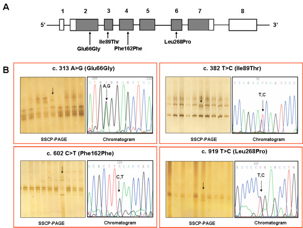
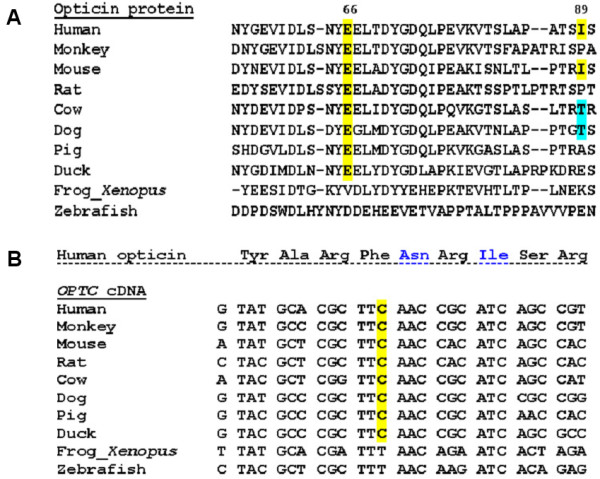
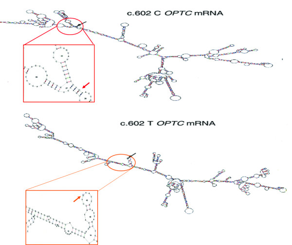
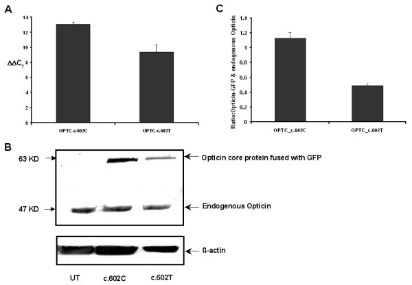
Results: We detected two missense (p.Glu66Gly & p.Ile89Thr) and one silent change (p.Phe162Phe; c.602 C>T) that was present in 3 different patients but in none of the 100 controls screened. The mutant (c.602T) mRNA was predicted to have remarkably different secondary structure compared to the wild-type transcript by in silico approaches. Subsequent wet-lab experiments showed lower expression of the gene both at the mRNA and protein levels.

Conclusion: Our study suggests OPTC as a candidate gene for POAG. Further, it highlights the importance of investigating the 'silent' variations for functional implication that might not be apparent from only in silico analysis.


Figures

Figure 1
Figure 1
Nucleotide variants in OPTC. (A): Gene structure of Opticin. Bars and lines represent the exons and introns, respectively. Exon numbers and mutation/polymorphism therein are indicated. Within exons shaded and open boxes show coding sequences and untranslated regions, respectively. (B): SSCP images along with chromatograms showing nucleotide changes in OPTC of POAG patients identified in Table 1.
Figure 2
Figure 2
Evolutionary conservation status of the silent change at cDNA level and other two nucleotide variants at protein level found in OPTC. (A): Multiple sequence alignment of opticin protein showing the conservation status of p.Glu66 (c.313A>G) residue and p.Ile89 (c.382T>C) residue across different vertebrate species. The sources of the sequences are NCBI: NP_055174.1 (human), XP_001102193.1 (monkey), NP_473417.1 (mouse), XP_222641.4 (rat), NP_991339.1 (cow), NP_001003056.1 (dog), NP_999173.1 (pig), NP_989804.1 (duck), NP_001016499.1 (Xenopus), ABB53599.1 (zebrafish). Except two lower vertebrates (Xenopus and zebrafish), p.66 (c.313A>G) is conserved in all higher vertebrate species, while p.Ile89 (c.382T>C) is conserved only in human and mouse. In both cases, multiple sequence alignment was done using ClustalW of EMBL-EBI [39] (B): Multiple sequence alignment of OPTC cDNA shows conservation of the wobble base for p.Phe162 which is altered (C>T) in case of a POAG patient (GL42 in Table 1). The sources of the sequences are GenBank: NM_014359 (human), XM_001102193.1 (monkey), NM_054076.1 (mouse), XM_001061460.1 (rat), NM_205770.1 (cow), NM_001003056.1 (dog), NM_214008.1 (pig), NM_204473.1 (duck), NM_001016499.2 (Xenopus) and NM_001003583.1 (zebrafish). Also, corresponding amino acid sequence of human opticin (NP_055174.1) is given above; except two lower vertebrates (Xenopus and zebrafish), c.602C (p.Phe162) is conserved in all higher vertebrate species.
Figure 3
Figure 3
Difference in the mRNA secondary structure between c.602C and c.602T variant in OPTC. Both the structures were predicted using the default parameter of RNAdraw (calculation temp. 37°C). Region of the mRNA harboring the variant nucleotide (indicated by arrow) has been enlarged for both the alleles to demonstrate the local structural alteration as predicted by RNAdraw.
Figure 4
Figure 4
Difference in mRNA and protein expression levels between c.602C and c.602T variants in OPTC. (A): Quantiative RT-PCR showing expression of c.602C and c.602T in mRNA level. The difference in mRNA expression is represented by ΔΔCT value. (B): Western blot of two variants of Opticin core protein fused with GFP as well as endogenous Opticin in RPE cells. UT represents the untransfected RPE cells. (C): Bar diagram showing the mean ± SD of densitometric scanning of western blots done thrice, that indicates much less expression of OPTC-c.602T variant (43%) than the normal one (OPTC-c.602C). Expression of transfected protein (Opticin-GFP) was normalized with endogenous Opticin.

References

    1. Libby RT, Gould DB, Anderson MG, John SW. Complex genetics of glaucoma susceptibility. Annu Rev Genomics Hum Genet. 2005;6:15–44. doi: 10.1146/annurev.genom.6.080604.162209. - DOI - PubMed
    1. Resnikoff S, Pascolini D, Etya'ale D, Kocur I, Pararajasegaram R, Pokharel GP, Mariotti SP. Global data on visual impairment in the year 2002. Bull World Health Organ. 2004;82:844–851. - PMC - PubMed
    1. Quigley HA. Number of people with glaucoma worldwide. Br J Ophthalmol. 1996;80:389–393. - PMC - PubMed
    1. Stone EM, Fingert JH, Alward WL, Nguyen TD, Polansky JR, Sunden SL, Nishimura D, Clark AF, Nystuen A, Nichols BE, Mackey DA, Ritch R, Kalenak JW, Craven ER, Sheffield VC. Identification of a gene that causes primary open angle glaucoma. Science. 1997;275:668–670. doi: 10.1126/science.275.5300.668. - DOI - PubMed
    1. Sarfarazi M, Child A, Stoilova D, Brice G, Desai T, Trifan OC, Poinoosawmy D, Crick RP. Localization of the fourth locus (GLC1E) for adult-onset primary open-angle glaucoma to the 10p15-p14 region. Am J Hum Genet. 1998;62:641–652. doi: 10.1086/301767. - DOI - PMC - PubMed

Publication types