Comparative Study
. 2007 May 1;305(1):187-201.
doi: 10.1016/j.ydbio.2007.02.004. Epub 2007 Feb 13.

Expanded and fat regulate growth and differentiation in the Drosophila eye through multiple signaling pathways

Affiliations
Comparative Study

Expanded and fat regulate growth and differentiation in the Drosophila eye through multiple signaling pathways

David M Tyler et al. Dev Biol. .

Abstract

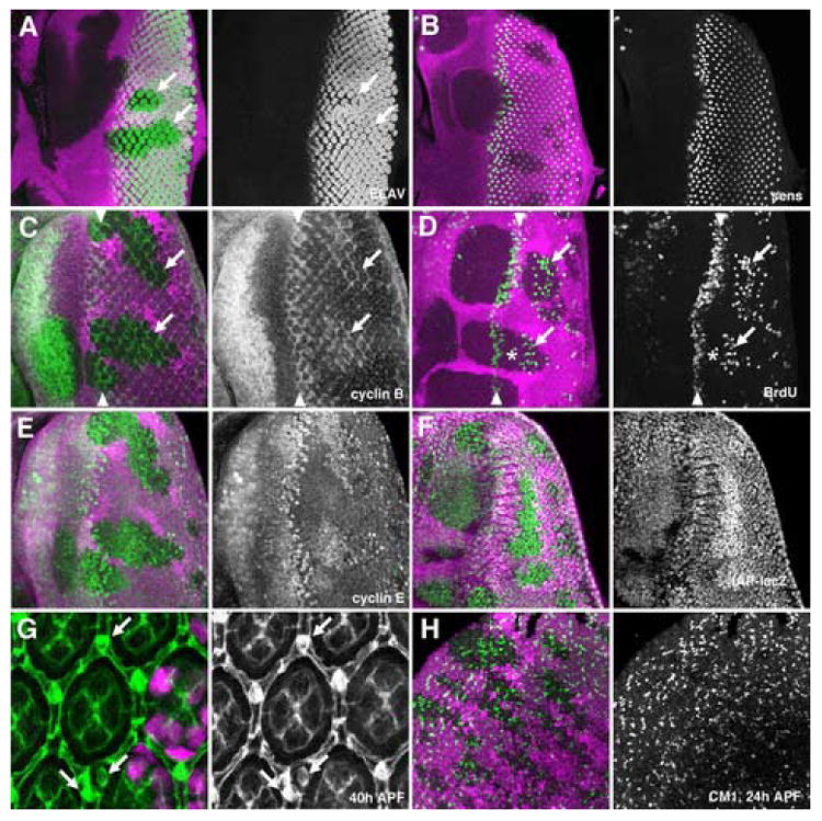
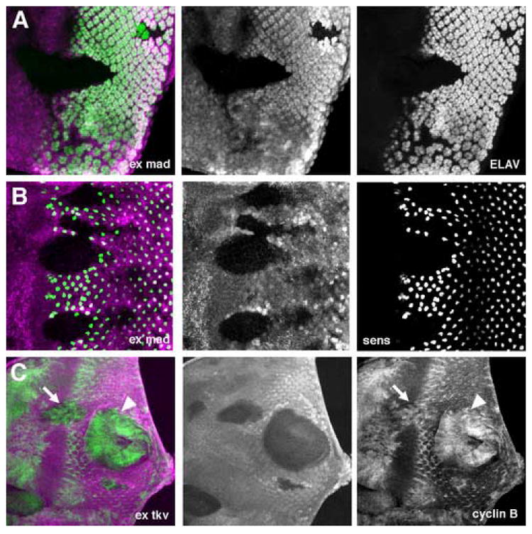
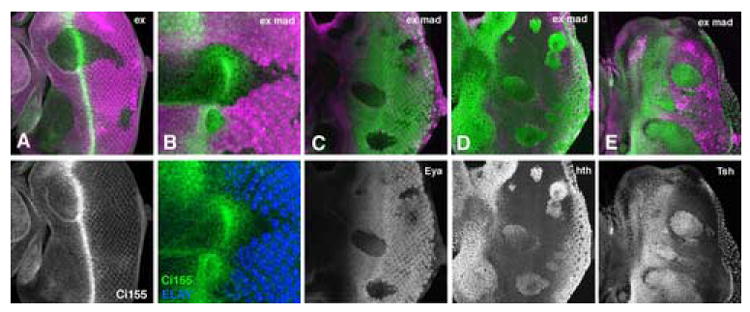
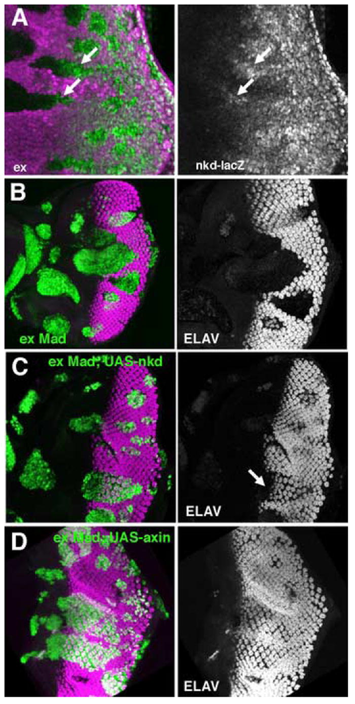
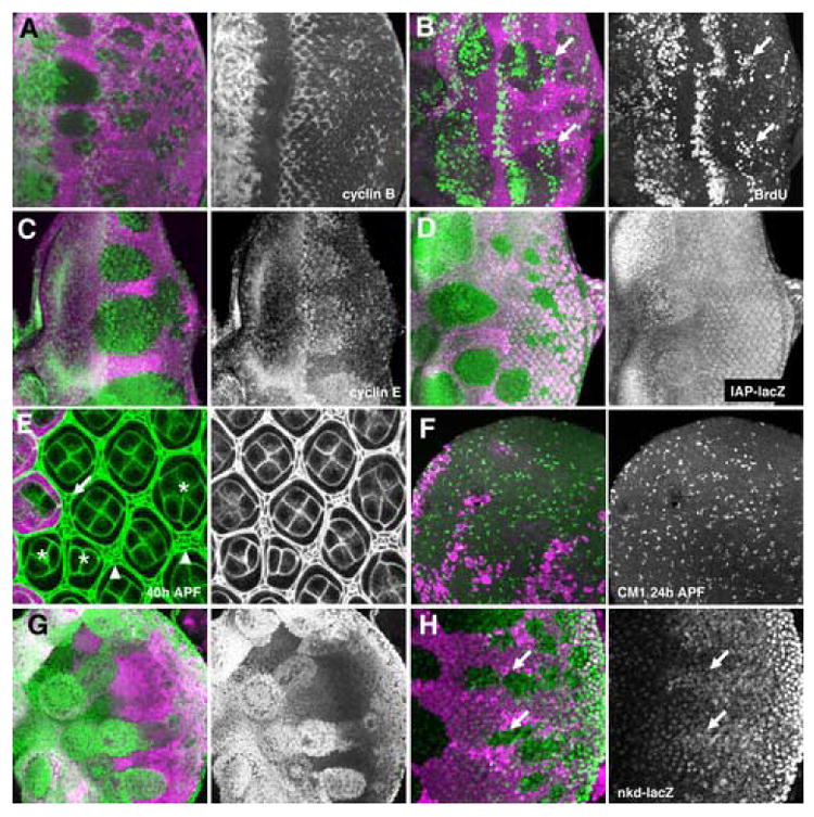
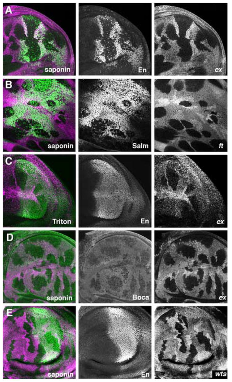
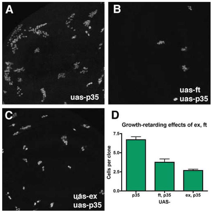
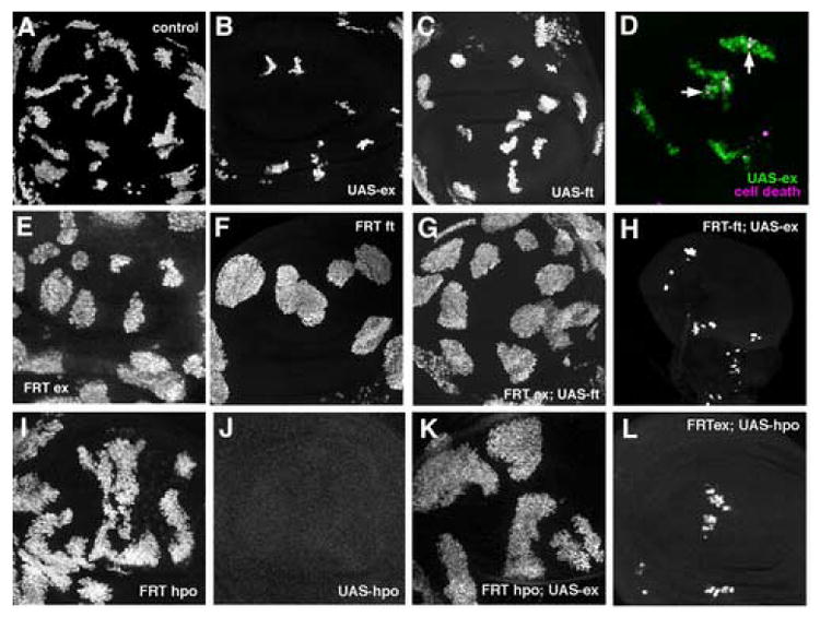
Mutations in the expanded gene act as hyperplastic tumor suppressors, interfere with cell competition and elevate Dpp signaling. Unlike Dpp overexpression, ex causes few patterning defects. Our data suggest that patterning effects are partly masked by antagonistic roles of other signaling pathways that are also activated. ex causes proliferation of cells in the posterior eye disc that are normally postmitotic. ex mutations elevate Wg signaling, but Dpp signaling antagonizes patterning effects of Wg. By contrast, if Dpp signaling is blocked in ex mutant cells, the elevated Wg signaling preserves an immature developmental state and prevents retinal differentiation. An effect of ex mutations on vesicle transport is suggested by evidence for altered sterol distribution. Mutations in ft show effects on proliferation, Wg signaling and sterols very similar to those of ex mutations. During disc growth, ex was largely epistatic to ft, and the Warts pathway mutation hippo largely epistatic to ex. Our data suggest that ft and ex act partially through the Warts pathway.


Figures

Figure 1
Figure 1. Differentiation and cell cycle in ex mutant clones
All figures show clones of homozygous ex mutant cells that lack the magenta lineage marker. Green labelling is ELAV (A), Sens (B), Cyclin B (C), BrdU (D), Cyclin E (E), IAP:LacZ (F), Discs Large (G), and CM1 (H). A) No difference in photoreceptor differentiation was visible between ex mutant and wild-type tissue. ELAV-expressing nuclei sometimes appear to be more widely-spaced in mutant tissue, perhaps because of the proliferation of intervening cells, but are the same size as in wild type. B) R8 photoreceptor differentiation occurred normally in ex mutant tissue, although a slight delay was sometimes apparent. C) Cyclin B expression marks cells in S-, G2-, and early M-phases of the cell cycle. Cycling cells can be seen at the SMW (arrowheads). In exNY1 mutant clones there are further interommatidial cells in the cell cycle iposterior to the SMW (arrows). D) S-phase cells are labeled in the SMW (arrowheads). Additional S-phases occured in exNY1 mutant clones (arrows). There was a gap (indicated by asterisk). between the normal S-phases of the SMW (asterisks) and later ectopic S-phases. E) Cyclin E protein levels were elevated in exNY1 mutant clones F) dIAP-LacZ transcription was elevated in exNY1 mutant clones G) anti-Discs large highlights cell membranes in pupal retina. The number and morphology of photoreceptor, cone pigment cells are the same in wild-type tissue and exNY1 mutant clones. There are ectopic bristle cells and duplicated bristles in ex mutant tissue (arrows). H) Similar amounts of pupal apoptosis occured in exNY1 mutant clones an in wild type retina.
Figure 2
Figure 2. Differentiation and cell cycle without ex and Dpp signaling
Clones of homozygous ex Mad mutant cells (A,B) or ex tkv mutant cells (C) lack the magenta lineage marker. Green labelling is ELAV (A), Sens (B), Cyclin B (C). A. Most exe1mad12 mutant cells fail to differentiate as photoreceptors. B. Most exe1mad12 mutant clones fail to specify R8 photoreceptors. C. exe1tkva12 double mutant cells fail to arrest in G1 anterior to the morphogenetic furrow (arrow) or in the posterior eye disc (arrowhead).
Figure 3
Figure 3. Eye specification in ex Mad mutant cells
Clones of homozygous ex (A) or ex Mad (B-E) mutant cells lack the magenta lineage marker. A. Ci155 accumulates almost normally in ex mutant clones (Ci155 protein in green). Thus, a wave of Hh signal transduction passes through ex mutant clones, as in wild type. B. A wave of Ci155 accumulation is somewhat delayed in ex Mad mutant clones compared to wild type (Ci155 protein in green). Thus, a wave of Hh signal transduction passes through ex Mad mutant clones. Delay may reflect the greater distance to photoreceptor cells that are the source of Hh secretion, as these differentiate only in wild type regions (ELAV protein labelling of photoreceptor cells shown in blue). Note that the distance between photoreceptor cells and peak Ci155 accumulation is similar in wild type and ex Mad mutant regions. Panel B is shown at 2x greater magnification than other panels. C. Eya protein (green) is not induced in exe1Mad12 mutant clones. Eya is necessary for eye specification. D. Hth protein (green) accumulates to high levels in exe1mad12 clones. E. Tsh protein (green) accumulates to high levels in exe1mad12 clones.
Figure 4
Figure 4. Elevated Wg signaling in ex mutant clones
In panel A, clones of ex mutant cells lack the magenta lineage marker. In panels B-C, clones of ex Mad mutant cells express GFP (Green). A. nkd-lacZ expression (green), a reporter of Wg signal transduction, is increased in exNY1 mutant clones. B. exe1mad12 clones marked by the expression of GFP (green) do not differentiate. Photoreceptor neurons labeled for ELAV (magenta). C. Differentiation is rescued by UAS-nkd:myc expression in exe1mad12 clones. Photoreceptor neurons labeled for ELAV (magenta). D. Dfferentiation is rescued in exe1mad12 clones.expressing UAS-axin. Photoreceptor neurons labeled for ELAV (magenta).
Figure 5
Figure 5. Differentiation, cell cycle, and eye specification in ft mutant clones
Clones of homozygous ft (A-F, H) or ft tkv (G) mutant cells lack the magenta lineage marker. A. ft mutant clones display similar cell cycle defects to ex. Cyclin B (green) is elevated in posterior cells in the eye disc. B. BrdU incorporation (green) shows ectopic S-phases in ft mutant clones, posterior to the furrow (arrow). C. Cyclin E protein (green) accumulates to higher levels in ft mutant clones. D. diap-lacZ expression (green) is elevated to a lesser degree in ftNY1 mutant clones than was seen for ex mutant clones (compare Figure 1F), and apparent only anterior to the morphogenetic furrow. E. Discs Large protein outlines cells in the pupal retina (green). ft mutant clones contain a few supernumary pigment cells(arrows), and ectopic and duplicated bristles (arrowheads). Some ft mutan ommatidia have abnormal numbers of cone cells; 2, 3 or 5 cells compared to 4 in wild-type (asterisks). F. Pupal apoptosis (CM1 antibody labelling in green) occurs normally in ft mutant clones. G. Hth protein expression (green) is maintained in ft tkv mutant clones. H. nkd-lacZ expression (green), a reporter of Wg signal transduction, is increased in ftNY1 mutant clones.
Figure 6
Figure 6. Detergent permeabilization of fixed cells
Clones of homozygous ft or ex mutant cells lack the magenta lineage marker in the wing disc. A. Nuclear En protein (green) is not detected in ex mutant cells permeabilized with saponin. Scattered mitotic cells are labeled within the clones, as expected from nuclear membrane breakdown. En is normally expressed in posterior compartments of the wing disc. B. Nuclear Salm protein (green) is not detected in ft mutant cells permeabilized with saponin. C. Nuclear En protein (green) is unaffected by ex mutations when Triton X-100 is used to permeabilize the preparation. D. The luminal ER protein Boca (green) is detected at reduced levels in ex mutant cells permeabilized with saponin. E. Nuclear En protein (green) is readily detected in wts mutant cells permabilized with saponin.
Figure 7
Figure 7. Growth inhibition by ectopic ex or ft
Panels A-C show clones of UAS:GFP UAS:p35-expressing cells induced by FLP-induced recombination to activate an Act:Gal4 transgene. B. Simultaneous overexpression of ft from a UAS:ft transgene reduces clonal growth 1.8-fold. C. Simultaneous overexpression of ex form aUAS:ex transgene reduces clonal growth 2.5-fold. D. Quantification of the results (p35, N=23; p35 ft, N=16; p35 ex, N=40).
Figure 8
Figure 8. Epistasis studies of ft , ex and hpo mutants and overexpression
MARCM was used to combine overexpression of GFP, and either ft ,ex, or hpo, with mitotic clones of mutant chromosomes in the wing imaginal disc. GFP expression marks the clones (green in panel B). A. Otherwise wild type, GFP-expressing control clones induced in parallel with panels C-H. B. ex overexpressing clones C. ft overexpressing clones D. Ex overexpression induced cell death both within and nearby the clones. Cell death identified by CM1 labelling (magenta). Dying, Ex-expressing cells appear white (eg vertical arrow); dying, non-expressing cells appear magenta (eg horizontal arrow). E. ex mutant clones. F. ft mutant clones G. ft overexpression in ex mutant clones. H. ex overexpression in ft mutant clones. Magnification is the same as other panels; small disc size seems to be a non-autonomous effect I. hpo mutant clones J. hpo overexpressing clones (clones were not recovered). K. ex overexpression in hpo mutant clones. L. hpo overexpression in ex mutant clones.

References

    1. Baker NE. Transcription of the segment polarity gene wingless in the imaginal discs of Drosophila, and the phenotype of a pupal-lethal wg mutation. Development. 1988;102:489–497. - PubMed
    1. Baker NE, Yu SY. The EGF receptor defines domains of cell cycle progression and survival to regulate cell number in the developing Drosophila eye. Cell. 2001;104:699–708. - PubMed
    1. Baonza A, Freeman M. Notch signalling and the initiation of neural development in the Drosophila eye. Development. 2001;128:3889–98. - PubMed
    1. Baonza A, Freeman M. Control of Drosophila eye specification by Wingless signalling. Development. 2002;129:5313–22. - PubMed
    1. Bennett FC, Harvey KF. Fat cadherin modulates organ size in Drosophila via the Salvador/Warts/Hippo signaling pathway. Curr Biol. 2006;16:2101–2110. - PubMed

Publication types

MeSH terms

LinkOut - more resources