. 2007 Apr 15;313(7):1326-36.
doi: 10.1016/j.yexcr.2007.02.001. Epub 2007 Feb 7.

Detyrosinated microtubule protrusions in suspended mammary epithelial cells promote reattachment

Affiliations

Detyrosinated microtubule protrusions in suspended mammary epithelial cells promote reattachment

Rebecca A Whipple et al. Exp Cell Res. .

Abstract

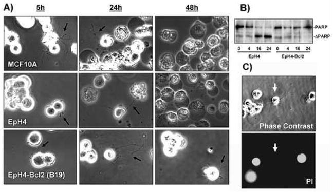
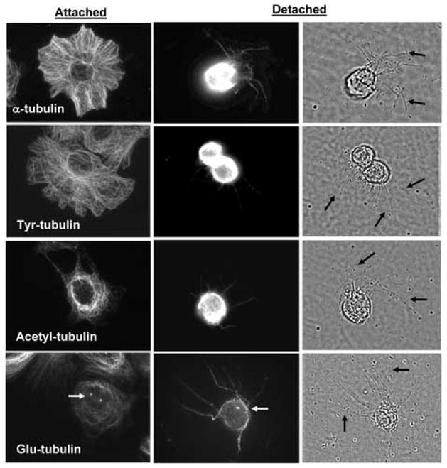
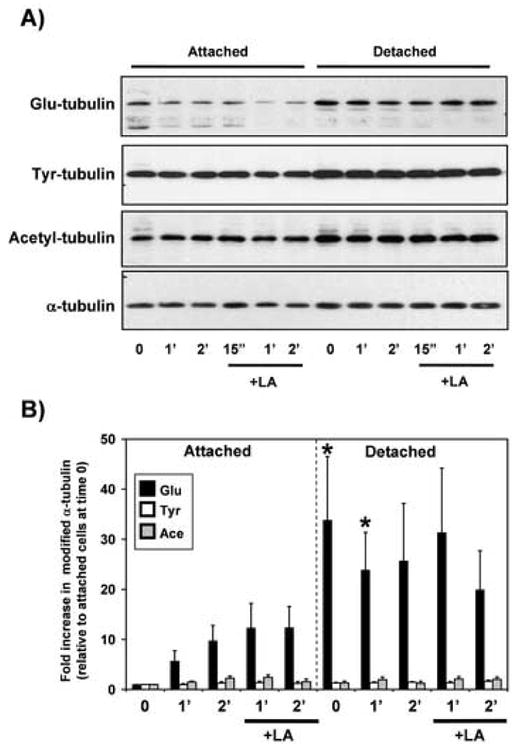
Breast tumor cells enter the bloodstream long before the development of clinically evident metastasis. However, the early presence of such bloodborne cells predicts poor patient outcome. Nearly 90% of human breast tumors arise as carcinomas from mammary epithelial cells, so it is important to study how these cells respond to the detached conditions that they would experience in the bloodstream. We report here that mammary epithelial cell lines produce long and dynamic protrusions of the plasma membrane when detached. Although human and mouse mammary epithelial cell lines die by apoptosis within 16 h of detachment, this protrusive response persists for days in cells overexpressing either Bcl-2 or Bcl-xL. Unlike actin-dependent invadopodia and podosomes, these protrusions are actually enhanced by actin depolymerization with Cytochalasin-D or Latrunculin-A. Immunofluorescence and Western blotting demonstrate that the protrusions are enriched in detyrosinated Glu-tubulin, a post-translationally modified form of alpha-tubulin that is found in stabilized microtubules. Video microscopy indicates that these protrusions promote cell-cell attachment, and inhibiting microtubule-based protrusions correlates with reduced extracellular matrix attachment. Since bloodborne metastasis depends on both cell-cell and cell-matrix attachment, microtubule-based protrusions in detached mammary epithelial cells provide a novel mechanism that could influence the metastatic spread of breast tumors.


Figures

Figure 1
Figure 1
Detachment induces cellular protrusions in both normal and Bcl2-expressing mammary epithelial cells. A) Human MCF10A or mouse EpH4 mammary epithelial cell lines produce protrusions of the plasma membrane when plated over agarose and photographed live (black arrows). Apoptosis becomes apparent in detached MCF10A and EpH4 cells after 24 hours of detachment and almost all cells are dead by 48 hours, as indicated by darkening and condensation of the cells. EpH4 cells that overexpress Bcl-2 (EpH4-Bcl2) remain largely viable at 48 hours and continue to produce protrusions. B) EpH4 cells or those overexpressing Bcl-2 were placed in suspension for the indicated time in hours. Cleavage of PARP to a truncated form (ΔPARP), indicates that apoptosis is largely complete in EpH4 cells after 16 hours, while relatively few EpH4-Bcl2 cells die even after 24 hours. C) EpH4 cells which produce protrusions (white arrows) continue to exclude propidium iodide, indicating an intact plasma membrane. Similar results were obtained with MCF10A cells.
Figure 2
Figure 2
Cytoskeletal inhibitors affect cellular protrusions. A) After 15 minutes of suspension, protrusions in EpH4 and MCF10A cells are quite small (Control, white arrows). Treatment with the actin depolymerizing agent, Cytochalasin-D, increased protrusions (white arrowheads) and this effect was even more pronounced with the actin inhibitor Latrunculin-A (LA, 5μM). The microtubule depolymerizing agent, Colchicine (1μM) prevented protrusions. The combination of LA and Colchicine (LA+Col) prevented protrusion formation, and those that did form appeared to be fragmented (black arrow). B) Populations of live, suspended cells were scored blindly for two or more protrusions longer than the cell radius. Each bar represents the mean +S.D. for three experiments in which at least 100 single cells were counted.
Figure 3
Figure 3
Protrusions are microtubule-based. EpH4 cells were suspended in either DMEM (Control) or media containing 5μM Latrunculin-A (LA), 1μM Colchicine or the combination of both drugs for 30 minutes. Cell were then spun onto glass coverslips and fluorescently stained for α-tubulin (green) and polymerized actin (red). Protrusions that are difficult to see by phase contrast are easily discernible with immunostaining for α-tubulin (white arrows). Protrusions contain little polymerized actin, except at points of concentration, which are often at the end (arrowhead). Depolymerization of actin with LA strongly enhances the formation of microtubule protrusions.
Figure 4
Figure 4
Protrusions are enriched in detyrosinated α-tubulin. EpH4-Bcl2 cells were grown on glass coverslips (Attached) or suspended over agarose for one hour in the presence of LA (5μM) to enhance protrusion formation (Detached). Immunostaining for different posttranslationally-modified forms of tubulin indicates differential localization in attached and suspended cells. Full-length α-tubulin, Tyr-tubulin and Acetyl-tubulin localize predominantly to the cell body in detached cells rather than specifically along the extended protrusions visible by phase-contrast (black arrows). Detyrosinated α-tubulin (Glu-tubulin) is enriched in the protrusions relative to the cell body, and is also found in centrosomes (white arrows).
Figure 5
Figure 5
Cellular levels of Glu-tubulin increase in response to detachment. EpH4-Bcl2 cells were grown attached to tissue culture dishes or suspended over agarose for the indicated times in either the absence or presence of LA (5μM). A) Cell lysates were immunoblotted to detect levels of modified tubulin isoforms in the cell populations. B) Modified α-tubulin isoforms were normalized to total α-tubulin and then compared relative to the levels in attached cells at time zero. Bars represent the mean + S.E.M. of four independent experiments. While Glu-tubulin increases significantly in response to detachment (P<0.05, t-test), none of the other tubulin forms increase significantly (P>0.05, t-test). Differences in Glu-tubulin upon detachment were also statistically-significant when compared to matched controls at time zero or one hour (black asterisks). LA treatment does not significantly increase cellular levels of Glu-tubulin in either attached or detached cells (P>0.05, t-test).
Figure 6
Figure 6
Microtubule protrusions are required for efficient cell attachment. A) EpH4-Bcl2 cells were suspended over agarose and treated with inhibitors as above. Following treatment, cells were plated into 0.3% methylcellulose media over agarose and the rate of cell-cell attachment was followed by Hoescht DNA staining. While LA-treated cells cluster similarly or even more tightly compared to control cells after 5 hours (black arrow), cells treated with tubulin polymerization inhibitors are signficantly delayed in clustering. Similar results were found in three independent experiments. B) EpH4-Bcl2 cells suspended over agarose for one hour in DMEM or DMEM containing LA (5μM), Colchicine (1μM) or the combination. Reattachment of these cells to either uncoated tissue culture plates or those pre-coated with laminin was followed by XTT cell viability assay. Values represent mean+S.D. of raw XTT values of three separate experiments. While actin depolymerization with LA does not affect cell-surface attachment, depolymerization of tubulin with colchicine significantly prevents initial cell attachment to either uncoated or laminin-coated plates. Additional time-lapse studies of protrusions during cell attachment are available as supplemental data.

References

    1. Naumov GN, MacDonald IC, Weinmeister PM, Kerkvliet N, Nadkarni KV, Wilson SM, Morris VL, Groom AC, Chambers AF. Persistence of solitary mammary carcinoma cells in a secondary site: a possible contributor to dormancy. Cancer Res. 2002;62:2162–8. - PubMed
    1. Naumov GN, MacDonald IC, Chambers AF, Groom AC. Solitary cancer cells as a possible source of tumour dormancy? Semin Cancer Biol. 2001;11:271–6. - PubMed
    1. Schmidt-Kittler O, Ragg T, Daskalakis A, Granzow M, Ahr A, Blankenstein TJ, Kaufmann M, Diebold J, Arnholdt H, Muller P, Bischoff J, Harich D, Schlimok G, Riethmuller G, Eils R, Klein CA. From latent disseminated cells to overt metastasis: genetic analysis of systemic breast cancer progression. Proc Natl Acad Sci U S A. 2003;100:7737–42. - PMC - PubMed
    1. Chambers AF, Groom AC, MacDonald IC. Dissemination and growth of cancer cells in metastatic sites. Nat Rev Cancer. 2002;2:563–72. - PubMed
    1. Frisch SM, Francis H. Disruption of epithelial cell-matrix interactions induces apoptosis. J Cell Biol. 1994;124:619–26. - PMC - PubMed

Publication types