. 2007 Mar 20;104(12):5067-72.
doi: 10.1073/pnas.0700929104. Epub 2007 Mar 14.

Human herpesvirus 6A accelerates AIDS progression in macaques

Affiliations

Human herpesvirus 6A accelerates AIDS progression in macaques

Paolo Lusso et al. Proc Natl Acad Sci U S A. .

Abstract

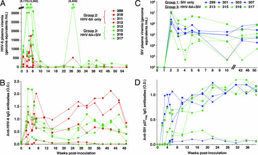
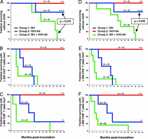
Although HIV is the necessary and sufficient causative agent of AIDS, genetic and environmental factors markedly influence the pace of disease progression. Clinical and experimental evidence suggests that human herpesvirus 6A (HHV-6A), a cytopathic T-lymphotropic DNA virus, fosters the progression to AIDS in synergy with HIV-1. In this study, we investigated the effect of coinfection with HHV-6A on the progression of simian immunodeficiency virus (SIV) disease in pig-tailed macaques (Macaca nemestrina). Inoculation of HHV-6A resulted in a rapid appearance of plasma viremia associated with transient clinical manifestations and followed by antibody seroconversion, indicating that this primate species is susceptible to HHV-6A infection. Whereas animals infected with HHV-6A alone did not show any long-term clinical and immunological sequelae, a progressive loss of CD4(+) T cells was observed in all of the macaques inoculated with SIV. However, progression to full-blown AIDS was dramatically accelerated by coinfection with HHV-6A. Rapid disease development in dually infected animals was heralded by an early depletion of both CD4(+) and CD8(+) T cells. These results provide in vivo evidence that HHV-6A may act as a promoting factor in AIDS progression.


Conflict of interest statement

The authors declare no conflict of interest.

Figures

Fig. 1.
Fig. 1.
Virological markers in pig-tailed macaques singly or dually infected with SIVsmE660 and HHV-6AGS. (A and B) Course of HHV-6A plasma viremia (A) and anti-HHV-6A IgG antibodies (B) during the acute phase of infection and the first year of follow-up in pig-tailed macaques singly infected with HHV-6A (group 2, red symbols) or coinfected with HHV-6A and SIV (group 3, green symbols). Time 0 corresponds to the time of HHV-6A inoculation; (C and D) Course of SIV plasma viremia (C) and anti-SIV p27Gag IgG antibodies (D) during the acute phase of infection and the first year of follow-up in pig-tailed macaques singly infected with SIV (group 1, blue symbols) or coinfected with HHV-6A and SIV (group 3, green symbols). Time 0 corresponds to the time of SIV inoculation. Coinfected monkeys were inoculated with HHV-6A at day 14 after SIV inoculation.
Fig. 2.
Fig. 2.
Clinical and immunological disease progression in pig-tailed macaques singly or dually infected with SIVsmE660 and HHV-6AGS. Shown are Kaplan–Meier curves for pig-tailed macaques singly infected with SIV (group 1, blue lines) or HHV-6A (group 2, red lines) or coinfected with SIV and HHV-6A (group 3, green lines). (A and D) AIDS-free survival. Survival analysis was performed nonparametrically by means of Kaplan–Meier curves. Log-rank tests were used to investigate differences in survival between groups. (B and E) Depletion of peripheral blood CD4+ T cells. (C and F) Depletion of peripheral blood CD8+ T cells. A–C shows Kaplan–Meier curves calculated for the original three groups of animals (group 3, n = 4). D–F shows Kaplan–Meier curves calculated after inclusion in group 3 of two additional animals (310 and 312), which were accidentally superinfected with SIV at month 13 and 21, respectively, from the initial HHV-6A inoculation (group 3, n = 6). For these two animals, survival analysis was performed starting from the time of SIV superinfection. The asterisks denote data that were censored because of drop-out or study termination. In group 2, only two animals (309 and 311) regularly completed the follow-up, because the remaining two animals were reclassified as SIV/HHV-6A coinfected during the course of the study.
Fig. 3.
Fig. 3.
Histopathology and in situ hybridization in lymph node tissues from macaques singly or dually infected with SIVsmE660 and HHV-6AGS. Tissues from two representative macaques are shown. Animal 303, singly infected with SIV (A and C); 317, coinfected with HHV-6A and SIV (B and D). A and B show Hematoxylin-eosin staining. C and D show In situ hybridization for SIV RNA of successive sections from the same lymph nodes as above. The arrows indicate enlarged lymphoid follicles with reactive germinal centers (A and B) and specific SIV RNA hybridization signal in correspondence to reactive germinal centers (C and D). In tissue from animal 303, the overall architecture is conserved, with lymphoid follicles barely discernible in the subcapsular area; low levels of SIV RNA are visible throughout the parenchyma, with little, if any, specific signal within reactive germinal centers. In tissue from animal 317, the histological architecture is largely effaced by a striking, florid follicular hyperplasia with clear-appearing, confluent reactive germinal centers; an intense SIV RNA signal is visible in correspondence to both subcapsular and deeper-located hyperplastic follicles.

Similar articles

Cited by

References

    1. Salahuddin SZ, Ablashi DV, Markham PD, Josephs SF, Sturzen-negger S, Kaplan M, Halligan G, Biberfeld P, Wong-Staal F, Kramarsky B, Gallo RC. Science. 1986;234:596–601. - PubMed
    1. Ablashi DV, Balachandran N, Josephs SF, Hung CL, Krueger GRF, Kramarski B, Salahuddin SZ, Gallo RC. Virology. 1991;184:545–552. - PubMed
    1. Yamanishi K, Okuna T, Shiraki K. Lancet. 1988;1:1065–1067. - PubMed
    1. Lusso P, Gallo RC. Immunol Today. 1995;16:67–71. - PubMed
    1. Lusso P, Markham PD, Tschachler E, DiMarzo Veronese F, Salahuddin SZ, Ablashi DV, Pahwa S, Gallo RC. J Exp Med. 1988;167:1659–1670. - PMC - PubMed

Publication types

MeSH terms