. 2007 Mar 20;104(12):5115-20.
doi: 10.1073/pnas.0700442104. Epub 2007 Mar 14.

Coxsackie B4 virus infection of beta cells and natural killer cell insulitis in recent-onset type 1 diabetic patients

Affiliations

Coxsackie B4 virus infection of beta cells and natural killer cell insulitis in recent-onset type 1 diabetic patients

Francesco Dotta et al. Proc Natl Acad Sci U S A. .

Abstract

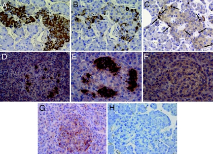
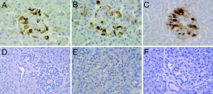
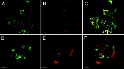
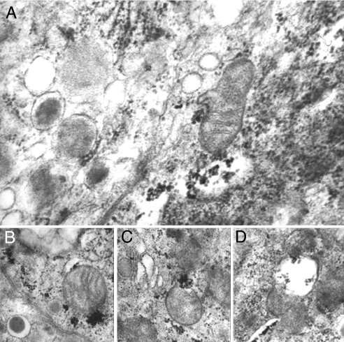
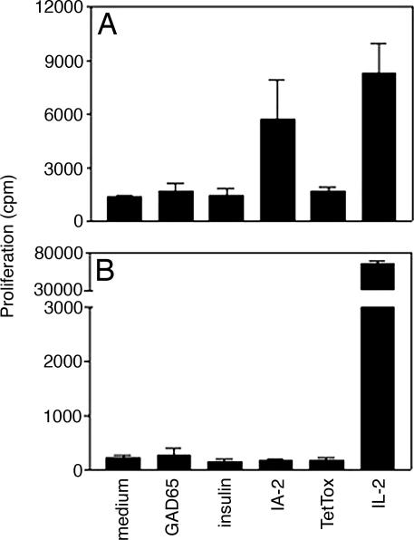
Type 1 diabetes is characterized by T cell-mediated autoimmune destruction of pancreatic beta cells. Several studies have suggested an association between Coxsackie enterovirus seroconversion and onset of disease. However, a direct link between beta cell viral infection and islet inflammation has not been established. We analyzed pancreatic tissue from six type 1 diabetic and 26 control organ donors. Immunohistochemical, electron microscopy, whole-genome ex vivo nucleotide sequencing, cell culture, and immunological studies demonstrated Coxsackie B4 enterovirus in specimens from three of the six diabetic patients. Infection was specific of beta cells, which showed nondestructive islet inflammation mediated mainly by natural killer cells. Islets from enterovirus-positive samples displayed reduced insulin secretion in response to glucose and other secretagogues. In addition, virus extracted from positive islets was able to infect beta cells from human islets of nondiabetic donors, causing viral inclusions and signs of pyknosis. None of the control organ donors showed signs of viral infection. These studies provide direct evidence that enterovirus can infect beta cells in patients with type 1 diabetes and that infection is associated with inflammation and functional impairment.


Conflict of interest statement

The authors declare no conflict of interest.

Figures

Fig. 1.
Fig. 1.
A type 1 diabetic pancreas shows nondestructive insulitis with NK cell infiltration and IFNα expression. (A–C) Immunohistochemistry panel showing reactivity for insulin (A), NK cells (B), and CD3+ cells (C) in consecutive sections representing the same islet; arrows point to scattered CD3+ cells (patient 1). (D–F) Reactivity to insulin in pancreatic sections from three additional type 1 diabetic patients (patients 4–6) showing a significant reduction of insulin-positive cells in these three individuals. Positivity for IFNα was detected in patient 1 (G) and in patients 2 and 3 (not shown), but not in control individuals (H). (Magnification: ×250.)
Fig. 2.
Fig. 2.
Reactivity to VP-1 enteroviral peptide in type 1 diabetic islet cells. (A–C) Immunohistochemical analysis shows reactivity to VP-1 enteroviral peptide in pancreatic islet cells from three different type 1 diabetic organ donors (patients 1–3). No VP-1 reactivity was observed in islets from another new-onset type 1 diabetic patient (D, patient 5) or in islets from control organ donors (E and F). (Magnification: ×250.)
Fig. 3.
Fig. 3.
Enteroviral infection affects only β cells in pancreatic islets. Confocal microscopy analysis demonstrated that β cells (A–C), but not α cells (D–F), result positive for VP-1 staining. Cells stained in green are positive for insulin (A and C) and glucagon (D and F), those stained in red are positive for VP-1 (B, E, and F), and those stained in yellow are double-positive cells for insulin and VP-1 (C). Double-positive cells for glucagon and VP-1 were not detected (F). (Scale bars: 18μm.)
Fig. 4.
Fig. 4.
Type 1 diabetic pancreas shows signs of viral infection. Electron microscopy of human islet β cells from a type 1 diabetic pancreas (patient 2): Many virus particles are present in the cytoplasm (A), frequently close to mitochondria (B and C) that sometimes appear swollen and partially destroyed (D). (Magnification: ×70,000.)
Fig. 5.
Fig. 5.
T cell autoreactivity is restricted to IA-2 and in peripheral lymphocytes only. Shown are T cell responses to various stimuli in PBMCs (A) and intraislet lymphocytes (B) recovered from islets isolated from the explanted pancreas allograft (patient 1).

References

    1. Gale EA. Diabetes. 2002;51:3353–3361. - PubMed
    1. Atkinson MA, Eisenbarth GS. Lancet. 2001;358:221–229. - PubMed
    1. von Herrath MG. Curr Top Microbiol Immunol. 2002;263:145–175. - PubMed
    1. Hyoty H, Hiltunen M, Lonnrot M. Clin Diagn Virol. 1998;9:77–84. - PubMed
    1. Yin H, Berg AK, Tuvemo T, Frisk G. Diabetes. 2002;51:1964–1971. - PubMed

Publication types

MeSH terms

Associated data