. 2007 Mar 20;104(12):4937-42.
doi: 10.1073/pnas.0700257104. Epub 2007 Mar 12.

Cytoskeletal dynamics of human erythrocyte

Affiliations

Cytoskeletal dynamics of human erythrocyte

Ju Li et al. Proc Natl Acad Sci U S A. .

Abstract

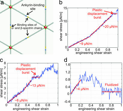
The human erythrocyte (red blood cell, RBC) demonstrates extraordinary ability to undergo reversible large deformation and fluidity. Such mechanical response cannot be consistently rationalized on the basis of fixed connectivity of the cell cytoskeleton that comprises the spectrin molecular network tethered to phospholipid membrane. Active topological remodeling of spectrin network has been postulated, although detailed models of such dynamic reorganization are presently unavailable. Here we present a coarse-grained cytoskeletal dynamics simulation with breakable protein associations to elucidate the roles of shear stress, specific chemical agents, and thermal fluctuations in cytoskeleton remodeling. We demonstrate a clear solid-to-fluid transition depending on the metabolic energy influx. The solid network's plastic deformation also manifests creep and yield regimes depending on the strain rate. This cytoskeletal dynamics model offers a means to resolve long-standing questions regarding the reference state used in RBC elasticity theory for determining the equilibrium shape and deformation response. In addition, the simulations offer mechanistic insights into the onset of plasticity and void percolation in cytoskeleton. These phenomena may have implication for RBC membrane loss and shape change in the context of hereditary hemolytic disorders such as spherocytosis and elliptocytosis.


Conflict of interest statement

The authors declare no conflict of interest.

Figures

Fig. 1.
Fig. 1.
An experimental in vitro demonstration of the “fluidization” of a healthy human RBC through a microfluidic channel at room temperature. The series of images show the shape of an RBC as it is squeezed through a 4 μm × 4 μm channel made of polydimethyloxysilane (PDMS), under a pressure differential of 1.5 mm of water. Note the recovery of shape upon egress from the channel. Images a, b, c, and d were taken at relative times of 0, 0.4, 0.8, and 1.4 s, respectively.
Fig. 2.
Fig. 2.
Coarse-grained cytoskeletal dynamics computer simulation. (a) Schematic of cytoskeleton of the human RBC. (b) A minimal physically realistic model with breakable actin–spectrin interaction. A (red sphere) represents an actin protofilament, and B (green and gray spheres) represents a spectrin segment. Only the two ending spectrin units (green spheres) of a spectrin chain can bind to A. (c) Interaction potentials between A–A (steric repulsion), A–Bend (Lennard–Jones potential), A–Bmid (steric repulsion), B–B[1] (spring potential between nearest neighbor spectrin units), and B–B[2] (steric repulsion between any other spectrin units). Bmid represents a nonending spectrin unit, and Bend represents an ending spectrin unit. Note that A–Bend forms breakable linkage. (d) Lipid membrane introduces additional effective repulsion between A–A (12), modeled by a linear potential with cutoff.
Fig. 3.
Fig. 3.
CD simulations with only mechanical energy input. (a) Snapshot of the equilibrium network structure at 300 K, top view and side view. (b) Snapshot of the sheared (γ = 1) network structure at 300 K, top view. (c) Shear stress-strain response at strain rate γ̇ = 3 × 10⁵ per s.
Fig. 4.
Fig. 4.
Shear stress-strain response of network at strain rate γ̇ = 3 × 10⁵ per s. (a) Energy hit rate 10 per μs and final network structure at γ = 1 (Inset). (b) Energy hit rate 2.5 per μs and final network structure at γ = 1 (Inset).
Fig. 5.
Fig. 5.
Refined CD simulation. (a) A model with breakable actin–spectrin and/or α-spectrin–β-spectrin interaction. Red spheres represent actin protofilaments, and green, gray, blue, and yellow spheres represent a spectrin segment. Only the two ending spectrin units (green spheres) of a spectrin chain can bind to red sphere. (b) Shear stress-strain response of network at γ̇ = 3 × 10⁵ per s and dimer–dimer association energy of 0.6 eV. Corresponding shear stress-strain response of network at 3 × 10⁵ per s and 0.56 eV (c) and 3 × 10⁵ per s and 0.47 eV (d) are shown.

Similar articles

Cited by

References

    1. Bennett V. Annu Rev Biochem. 1985;54:273–304. - PubMed
    1. Mohandas N, Evans E. Annu Rev Biophys Biomol Struct. 1994;23:787–818. - PubMed
    1. An XL, Lecomte MC, Chasis JA, Mohandas N, Gratzer W. J Biol Chem. 2002;277:31796–31800. - PubMed
    1. Cloitre M, Borrega R, Leibler L. Phys Rev Lett. 2000;85:4819–4822. - PubMed
    1. Bursac P, Lenormand G, Fabry B, Oliver M, Weitz DA, Viasnoff V, Butler JP, Fredberg JJ. Nat Mater. 2005;4:557–561. - PubMed

Publication types

LinkOut - more resources