. 2007 Mar 20;104(12):4949-54.
doi: 10.1073/pnas.0611640104. Epub 2007 Mar 14.

A lamin A protein isoform overexpressed in Hutchinson-Gilford progeria syndrome interferes with mitosis in progeria and normal cells

Affiliations

A lamin A protein isoform overexpressed in Hutchinson-Gilford progeria syndrome interferes with mitosis in progeria and normal cells

Kan Cao et al. Proc Natl Acad Sci U S A. .

Abstract

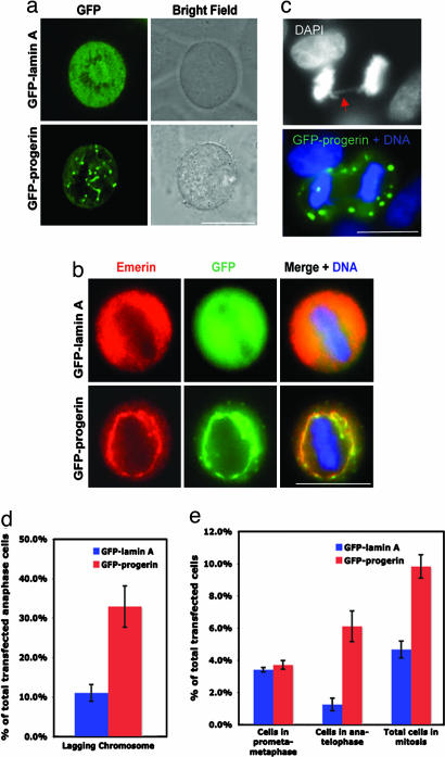
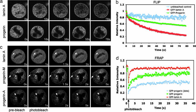
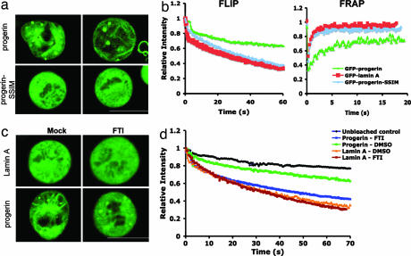
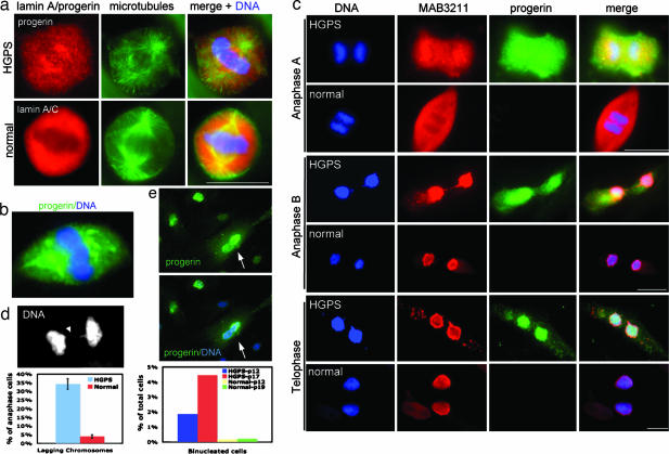
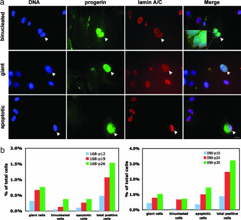
Hutchinson-Gilford progeria syndrome (HGPS) is a rare genetic disorder characterized by dramatic premature aging. Classic HGPS is caused by a de novo point mutation in exon 11 (residue 1824, C --> T) of the LMNA gene, activating a cryptic splice donor and resulting in a mutant lamin A (LA) protein termed "progerin/LADelta50" that lacks the normal cleavage site to remove a C-terminal farnesyl group. During interphase, irreversibly farnesylated progerin/LADelta50 anchors to the nuclear membrane and causes characteristic nuclear blebbing. Progerin/LADelta50's localization and behavior during mitosis, however, are completely unknown. Here, we report that progerin/LADelta50 mislocalizes into insoluble cytoplasmic aggregates and membranes during mitosis and causes abnormal chromosome segregation and binucleation. These phenotypes are largely rescued with either farnesyltransferase inhibitors or a farnesylation-incompetent mutant progerin/LADelta50. Furthermore, we demonstrate that small amounts of progerin/LADelta50 exist in normal fibroblasts, and a significant percentage of these progerin/LADelta50-expressing normal cells are binucleated, implicating progerin/LADelta50 as causing similar mitotic defects in the normal aging process. Our findings present evidence of mitotic abnormality in HGPS and may shed light on the general phenomenon of aging.


Conflict of interest statement

The authors declare no conflict of interest.

Figures

Fig. 1.
Fig. 1.
Mitotic defects in GFP-progerin/LAΔ50-transfected cells (progerin/LAΔ50 is referred to as progerin in all figures). (a) Live cell images of GFP-LA- and GFP-progerin-transfected mitotic cells. (b) Immunofluorescence on GFP-LA- or GFP-progerin-transfected mitotic cells, using an anti-emerin antibody. (c) DAPI staining showing a lagging chromosome (red arrow) in a GFP-progerin-transfected anaphase cell. (d) Quantification of abnormal chromosome segregation in GFP-LA- or GFP-progerin-transfected cells. (e) Quantification of GFP-LA- or GFP-progerin-transfected cells in early (from prometaphase to metaphase) or late (from anaphase to telophase) mitosis. A significant increase of cells in late mitosis was observed when GFP-progerin was transfected. (Scale bars: 20 μm.)
Fig. 2.
Fig. 2.
Abnormal dynamic behavior of GFP-progerin/LAΔ50 during mitosis. (a and b) FLIP analysis. (a) Selected images of GFP-LA- or GFP-progerin/LAΔ50-transfected mitotic cells during FLIP of the indicated area (yellow square). (b) Kinetics of overall FLIP (white circled area) in GFP-LA- or GFP-progerin/LAΔ50-transfected mitotic cells. (c and d) FRAP analysis. (c) Selected images of the GFP-progerin/LAΔ50- or GFP-LA-transfected mitotic cells during FRAP of the indicated area in the cytoplasm (as indicated by white arrows). Progerin-M, membrane-associated progerin/LAΔ50; progerin-A, aggregated progerin/LAΔ50 (bright dots). (d) GFP-LA or GFP-progerin/LAΔ50 FRAP kinetics in transfected mitotic cells. Values in b and d represent means from at least six different cells. Photographs were taken at ×60 magnification.
Fig. 3.
Fig. 3.
Retention of farnesylated C terminus causes the abnormal localization and dynamic behavior of GFP-progerin/LAΔ50 during mitosis. (a) Live cell images of GFP-progerin- and GFP-progerin-SSIM-transfected mitotic cells. Two examples are shown for each type. (b) Kinetics of overall FLIP (Left) and FRAP (Right) in GFP-LA-, GFP-progerin-, and GFP-progerin-SSIM-transfected mitotic cells. (c) Live cell images of GFP-LA- or GFP-progerin-transfected mitotic cells after mock (DMSO) or FTI (2.0 μM lonafarnib) treatment for 48 h. (d) Kinetics of overall FLIP in the mitotic cells in c. Values in b and d represent means from at least five different cells. (Scale bars: 20 μm.)
Fig. 4.
Fig. 4.
Mitotic defects in HGPS fibroblast cells. (a) Immunofluorescence on primary dermal fibroblasts from a normal father (HGADFN168) and a child with classic HGPS (HGADFN167) with anti-LA/lamin C or anti-progerin, respectively (red). The same cells were also counterstained with anti-α-tubulin (green) and DAPI for DNA (blue). (b) An example showing that anti-progerin antibody sometimes labeled large membrane-like aggregates in HGPS mitotic cells. (c) Double immunodetection with anti-progerin (green) and anti-LA/lamin C (red) antibodies on mitotic fibroblasts from a normal father (HGADFN168) and a child with classic HGPS (HGADFN167), showing delay of reassembly of progerin/LAΔ50 to the NE in late mitosis. (d) A representative image and quantification of abnormal chromosome segregation in HGPS and normal fibroblasts. Arrowhead points to a lagging chromosome. (e) Images and quantification of binucleation in the HGPS fibroblasts. Arrows point to a binucleated cell. (Scale bars: 20 μm.)
Fig. 5.
Fig. 5.
Expression of progerin/LAΔ50 in normal human fibroblasts. (a) Immunofluorescence of fibroblasts from an unaffected individual (HGADFN168), using anti-LA/lamin C (red) and anti-progerin (green). DNA is counterstained in blue with DAPI. Arrows indicate the progerin/LAΔ50-expressing normal cells. (Scale bar: 20 μm.) (b) Quantification of progerin/LAΔ50-positive normal cells in two independent normal fibroblast cell lines at various passages (HGADFN168 at passages 12, 19, and 26; HGADFN090 at passages 10, 24, and 30).

Similar articles

Cited by

References

    1. Gruenbaum Y, Margalit A, Goldman RD, Shumaker DK, Wilson KL. Nat Rev Mol Cell Biol. 2005;6:21–31. - PubMed
    1. Goldman RD, Gruenbaum Y, Moir RD, Shumaker DK, Spann TP. Genes Dev. 2002;16:533–547. - PubMed
    1. Sinensky M, Fantle K, Trujillo M, McLain T, Kupfer A, Dalton M. J Cell Sci. 1994;107:61–67. - PubMed
    1. Capell B, Collins F. Nat Rev Genet. 2006;7:940–952. - PubMed
    1. DeBusk FL. J Pediatr. 1972;80:697–724. - PubMed

Publication types