. 2007 Mar 27;104(13):5533-8.
doi: 10.1073/pnas.0611482104. Epub 2007 Mar 2.

Sensitivity of T cells to antigen and antagonism emerges from differential regulation of the same molecular signaling module

Affiliations

Sensitivity of T cells to antigen and antagonism emerges from differential regulation of the same molecular signaling module

Dennis C Wylie et al. Proc Natl Acad Sci U S A. .

Abstract

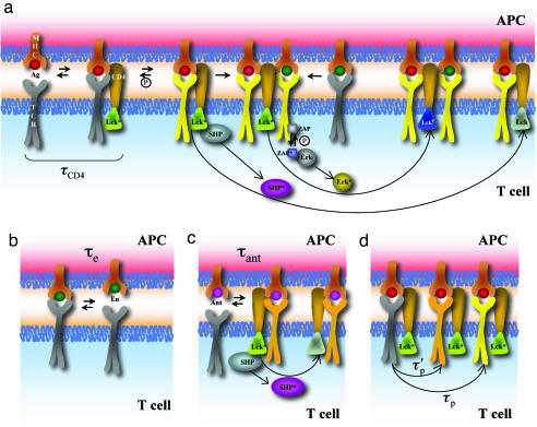
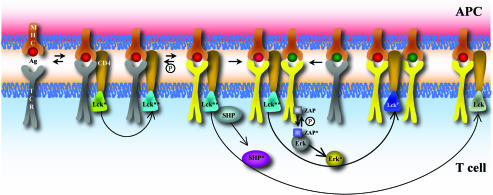
Activation of T helper cells is necessary for the adaptive immune response to pathogens, and spurious activation can result in organ-specific autoimmunity (e.g., multiple sclerosis). T cell activation is initiated by membrane-proximal signaling that is predicated on the binding of the T cell receptor expressed on the T cell surface to peptide major histocompatibility complex (pMHC) molecules presented on the surface of antigen-presenting cells. These signaling processes regulate diverse outcomes, such as the ability of T cells to discriminate sensitively between stimulatory pMHC molecules and those that are characteristic of "self," and the phenomenon of antagonism (wherein the presence of certain pMHC molecules impairs T cell receptor signaling). We describe a molecular model for membrane-proximal signaling in T cells from which these disparate observations emerge as two sides of the same coin. This development of a unified mechanism that is consistent with diverse data would not have been possible without explicit consideration of the stochastic nature of the pertinent biochemical events. Our studies also reveal that certain previously proposed concepts are not dueling ideas but rather are different stimuli-dependent manifestations of a unified molecular model for membrane-proximal signaling. This model may provide a conceptual framework for further investigations of early events that regulate T cell activation in response to self and foreign antigens and for the development of intervention protocols to inhibit aberrant signaling.


Conflict of interest statement

The authors declare no conflict of interest.

Figures

Fig. 1.
Fig. 1.
Schematic representation of the bare model. (a) Cooperative interactions between agonist and endogenous ligands in phosphorylating TCRs (1, 3, 16) and feedback regulation of Lck (12, 19). Agonist peptides (red) bind TCRs (gray) for a sufficiently long time to recruit Lck (dark green) in a time scale equal to τCD4. Lck can phosphorylate TCRs (yellow represents such TCRs). Fully phosphorylated TCRs bind ZAP70 (light blue) with high efficiency, and Lck can phosphorylate ZAP70 (dark blue represents activated ZAP70), which in turn results in ERK activation (ERK → ERK*). Endogenous peptides (green) can associate with signaling complexes nucleated by agonists, and TCRs that bind to these endogenous pMHC molecules can be phosphorylated by the proximal Lck. Lck can activate the phosphatase SHP (purple, when activated), which can deactivate Lck (light green represents its deactivated state). ERK* can phosphorylate Lck, and SHP cannot act on this form of Lck (dark blue). (b) TCR–endogenous pMHC complexes are extremely short-lived (with a lifetime of τe) and thus rarely recruit Lck. (c) As described in text, we hypothesize that the short-lived antagonist (lifetime τant) pMHC–TCR can recruit Lck but cannot carry out full phosphorylation of TCRs or nucleate signaling complexes as agonists can. The recruited Lck could activate SHP. (d) Agonist pMHC–TCR complexes recruit Lck, which phosphorylates the ITAMs partially or fully with the time scales τ′p and τp, respectively.
Fig. 2.
Fig. 2.
Results for the bare model. (a) The ratio of fully to partially phosphorylated TCRs in the bare model at time t = 250 s increases slightly as the number of antagonist pMHC ligands present is increased. There are 100 agonists and 5,000 endogenous pMHC. The other parameters used, and sensitivity (or lack thereof) of the results to variation in these parameters, are described in SI Appendix. (b) ERK activation increases with antagonist number past a certain threshold (between 200 and 500 antagonist pMHC). The parameters are the same as in a. (c) The increase in ERK activation with inclusion of “antagonist” pMHC ligands is robust over rates of antagonist dissociation from TCRs varying from 0.1 s−1 to 10 s−1. Filled diamonds are for 100 agonists and 5,000 endogenous pMHC; open squares include an additional 500 antagonists. The vertical dotted gray line indicates the very narrow window in which Eq. 4 is satisfied (τant from Eq. 2 is equal to the inverse of the dissociation rate on the x axis).
Fig. 3.
Fig. 3.
Results for the unified model. (a) The ratio of fully to partially phosphorylated TCRs in the unified model at time t = 250 s decreases with increasing numbers of antagonist pMHC. (b) ERK activation is suppressed by antagonist ligands in the unified model. (c) Phenomenon of antagonism in the unified model depends on antagonist pMHC–TCR dissociation rate. Antagonism is maximized (i.e., ERK activation is minimized) at an intermediate value of pMHC–TCR dissociation rate.
Fig. 4.
Fig. 4.
Schematic representation of the unified model. The colors and symbols are exactly the same as in Fig. 1a, except that Lck can potentially be converted to a state of higher activity (teal) if a TCR is bound to the corresponding pMHC for a sufficiently long time (details in text).

Similar articles

Cited by

References

    1. Irvine DJ, Purbhoo MA, Krogsgaard M, Davis MM. Nature. 2002;419:845–849. - PubMed
    1. Purbhoo MA, Irvine DJ, Huppa JB, Davis MM. Nat Immunol. 2004;5:524–530. - PubMed
    1. Krogsgaard M, Li QJ, Sumen C, Huppa JB, Huse M, Davis MM. Nature. 2005;434:238–243. - PubMed
    1. Grakoui A, Bromley SK, Sumen C, Davis MM, Shaw AS, Allen PM, Dustin ML. Science. 1999;285:221–227. - PubMed
    1. Monks CR, Freiberg BA, Kupfer H, Sciaky N, Kupfer A. Nature. 1998;395:82–86. - PubMed

Publication types