. 2007 Mar 20;104(12):5217-22.
doi: 10.1073/pnas.0609656104. Epub 2007 Mar 14.

Genetic deletion of p66(Shc) adaptor protein prevents hyperglycemia-induced endothelial dysfunction and oxidative stress

Affiliations

Genetic deletion of p66(Shc) adaptor protein prevents hyperglycemia-induced endothelial dysfunction and oxidative stress

Giovanni G Camici et al. Proc Natl Acad Sci U S A. .

Abstract

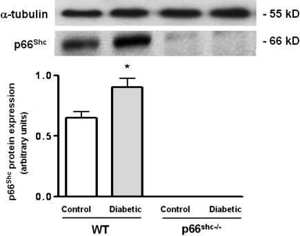
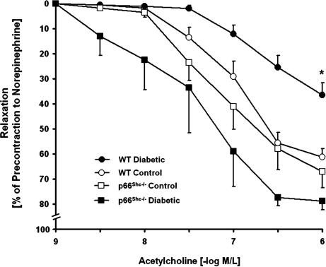
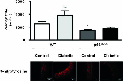
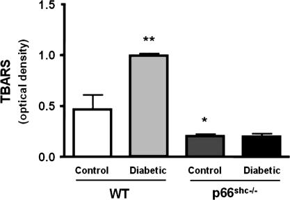
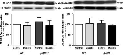
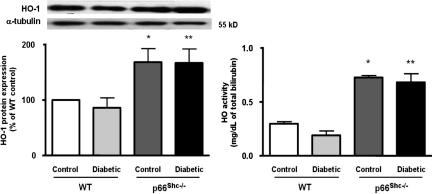
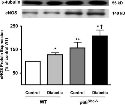
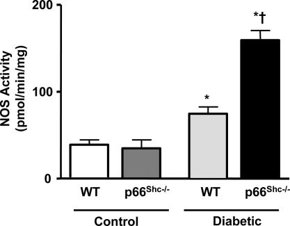
Increased production of reactive oxygen species (ROS) and loss of endothelial NO bioavailability are key features of vascular disease in diabetes mellitus. The p66(Shc) adaptor protein controls cellular responses to oxidative stress. Mice lacking p66(Shc) (p66(Shc-/-)) have increased resistance to ROS and prolonged life span. The present work was designed to investigate hyperglycemia-associated changes in endothelial function in a model of insulin-dependent diabetes mellitus p66(Shc-/-) mouse. p66(Shc-/-) and wild-type (WT) mice were injected with citrate buffer (control) or made diabetic by an i.p. injection of 200 mg of streptozotocin per kg of body weight. Streptozotocin-treated p66(Shc-/-) and WT mice showed a similar increase in blood glucose. However, significant differences arose with respect to endothelial dysfunction and oxidative stress. WT diabetic mice displayed marked impairment of endothelium-dependent relaxations, increased peroxynitrite (ONOO(-)) generation, nitrotyrosine expression, and lipid peroxidation as measured in the aortic tissue. In contrast, p66(Shc-/-) diabetic mice did not develop these high-glucose-mediated abnormalities. Furthermore, protein expression of the antioxidant enzyme heme oxygenase 1 and endothelial NO synthase were up-regulated in p66(Shc-/-) but not in WT mice. We report that p66(Shc-/-) mice are resistant to hyperglycemia-induced, ROS-dependent endothelial dysfunction. These data suggest that p66(Shc) adaptor protein is part of a signal transduction pathway relevant to hyperglycemia vascular damage and, hence, may represent a novel therapeutic target against diabetic vascular complications.


Conflict of interest statement

The authors declare no conflict of interest.

Figures

Fig. 1.
Fig. 1.
p66Shc protein expression in aortic lysates from WT control and diabetic mice. Bar graphs show densitometric analysis of Western blot of p66Shc protein in control and diabetic WT and p66Shc−/− mice. α-Tubulin was used as a reference protein. Data are presented as mean ± SEM; n = 3 in each group. ∗, P < 0.05 vs. control WT mice.
Fig. 2.
Fig. 2.
Isometric tension studies in aortic rings from control and diabetic WT and p66Shc−/− mice. Vessel relaxations to the endothelium-dependent agonist acetylcholine were normal in control WT (open circles) and p66Shc−/− (open squares) mice. Diabetic WT mice (filled circles) exhibited impaired endothelium-dependent relaxations compared with diabetic p66Shc−/− mice (filled squares) and with control WT mice (open circles). There were no differences in endothelium-dependent relaxations between diabetic and control p66Shc−/− mice. Relaxations were obtained during contractions to 10 −6 mol/liter norepinephrine. Data are presented as mean ± SEM; n = 6–9 in each group. ∗, P < 0.05 for diabetic WT mice vs. all other groups.
Fig. 3.
Fig. 3.
Bar graphs show ONOO levels determined by chemiluminescence. Results are presented as mean ± SEM; n = 6 in each group. ∗, P < 0.05 vs. control WT mice; ∗∗, P < 0.05 vs. control WT mice. Immunostaining of nitrotyrosine residues in aortas from control and diabetic WT and p66Shc−/− mice is shown.
Fig. 4.
Fig. 4.
TBARS levels. Bar graphs show TBARS levels in aortas from control and diabetic WT and p66Shc−/− mice. Results are presented as mean ± SEM; n = 4 in each group. ∗, P < 0.05 vs. control WT mice; ∗∗, P < 0.05 vs. control WT.
Fig. 5.
Fig. 5.
Protein expression from aortas of control and diabetic WT and p66Shc−/− mice. (Left) MnSOD. (Right) Cu/ZnSOD. Bar graphs show densitometric analysis of Western blots of MnSOD and Cu/ZnSOD in control and diabetic WT and p66Shc−/− mice, respectively. Results are presented as mean ± SEM; n = 4–6 in each group.
Fig. 6.
Fig. 6.
HO-1 protein expression and total HO activity from aortas of control and diabetic WT and p66Shc−/− mice. (Left) HO-1 protein expression from aortas of control and diabetic WT and p66Shc−/− mice. Bar graphs show densitometric analysis of Western blots of HO-1 protein in control and diabetic WT and p66Shc−/− mice. (Right) HO activity in control and diabetic WT and p66Shc−/− mice. Results are presented as mean ± SEM; n = 4 in each group. ∗, P < 0.05 vs. control WT mice; ∗∗, P < 0.05 vs. diabetic WT mice.
Fig. 7.
Fig. 7.
eNOS protein expression from aortas of control and diabetic WT and p66Shc−/− mice. Bar graphs show densitometric analysis of Western blots of eNOS protein in control and diabetic WT and p66Shc−/− mice. Results are presented as mean ± SEM; n = 6–9 in each group. ∗, P < 0.05 vs. respective control; ∗∗, P < 0.05 vs. control WT mice; †, P < 0.05 vs. diabetic WT mice.
Fig. 8.
Fig. 8.
NOS activity in WT and p66Shc−/− mice under control and diabetic conditions. Results are presented as mean ± SEM; n = 6 in each group. ∗, P < 0.05 vs. respective control; †, P < 0.05 vs. diabetic WT mice.

References

    1. Creager MA, Luscher TF, Cosentino F, Beckman JA. Circulation. 2003;108:1527–1532. - PubMed
    1. Widlansky ME, Gokce N, Keaney JF, Jr, Vita JA. J Am Coll Cardiol. 2003;42:1149–1160. - PubMed
    1. Lerman A, Zeiher AM. Circulation. 2005;111:363–368. - PubMed
    1. van der Loo B, Labugger R, Skepper JN, Bachschmid M, Kilo J, Powell JM, Palacios-Callender M, Erusalimsky JD, Quaschning T, Malinski T, et al. J Exp Med. 2000;192:1731–1744. - PMC - PubMed
    1. Turko IV, Murad F. Pharmacol Rev. 2002;54:619–634. - PubMed

Publication types

MeSH terms

Substances