. 2007 Mar 20;104(12):5109-14.
doi: 10.1073/pnas.0609611104. Epub 2007 Mar 12.

Hypoxia-inducible factor 1alpha stabilization by carbon monoxide results in cytoprotective preconditioning

Affiliations

Hypoxia-inducible factor 1alpha stabilization by carbon monoxide results in cytoprotective preconditioning

Beek Y Chin et al. Proc Natl Acad Sci U S A. .

Abstract

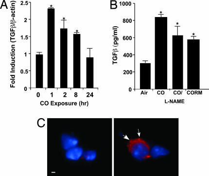
The most salient feature of carbon monoxide (CO)-mediated cytoprotection is the suppression of inflammation and cell death. One of the important cellular targets of CO is the macrophage (mphi). Many studies have shown that exposure of mphi to CO results in the generation of an antiinflammatory phenotype; however, these reports have ignored the effect of CO alone on the cell before stimulation. Most investigations have focused on the actions of CO in modulating the response to noxious stimuli. We demonstrate here that exposure of mphi to CO results in a significant and transient burst of reactive oxygen species (ROS) arising from the mitochondria (mitochondria-deficient mphi do not respond to CO to produce ROS). The ROS promote rapid activation and stabilization of the transcription factor hypoxia-inducible factor 1alpha (HIF-1alpha), which regulates expression of genes involved in inflammation, metabolism, and cell survival. The increase in HIF-1alpha expression induced by CO results in regulated expression of TGF-beta, a potent antiinflammatory cytokine. CO-induced HIF-1alpha and TGF-beta expression are necessary to prevent anoxia/reoxygenation-induced apoptosis in mphi. Furthermore, blockade of HIF-1alpha using RNA interference and HIF-1alpha-cre-lox mphi resulted in a loss of TGF-beta expression and CO-induced protection. A similar mechanism of CO-induced protection was operational in vivo to protect against lung ischemia-reperfusion injury. Taken together, we conclude that CO conditions the mphi via a HIF-1alpha and TGF-beta-dependent mechanism and we elucidate the earliest events in mphi signaling that lead to and preserve cellular homeostasis at the site of injury.


Conflict of interest statement

Conflict of interest statement: L.E.O. is a paid consultant of Linde Healthcare.

Figures

Fig. 1.
Fig. 1.
Induction of HIF-1α expression and activity after administration of CO. (A) Kinetics of HIF-1α protein via Western blot analyses, in mφ exposed to CO or a CORM. α-Tubulin and β-actin were used as loading controls. (B) Detection of HIF-1α DNA binding by EMSA. (C) HIF-1α luciferase reporter activity in indicated groups. ∗, P < 0.01 vs. control; ∗#, P < 0.05 vs. CO. (D) Immunofluorescent detection of cytoplasmic and nuclear HIF-1α (green punctate dots, denoted by arrows) in situ in 3D-reconstructed isolated lung mφ (air control, Upper; panel-CO exposed, Lower). Radiographs and luciferase data are representative of three to five independent experiments. Images are representative of 10 fields of view from air- and CO-exposed animals (n = 3 each). (Scale bar: 1 μm.)
Fig. 2.
Fig. 2.
Role of mitochondria in CO-induced HIF-1α expression. (A) Mitochondria-deficient (ρ°) and WT mφs were characterized by their ability to generate ROS by 2′,7′-dichlorodihydrofluorescein diacetate (DCF) fluorescence. (Upper) WT. (Lower) ρ°. Cells were preloaded with DCF ± CO, and fluorescence was determined by FACS (filled, 0 min; unfilled dashed line, 5 min; unfilled bold line, 60 min). (B) Western blot showing kinetics of HIF-1α protein in ρ° cells exposed to CO; α-tubulin is shown as a loading control. (C) Immunofluorescent staining for HIF-1α (red, denoted by arrows) in cultured WT mφs (left-air and right-CO; Upper) and ρ° cells (left-air and right-CO; Lower). Images shown are representative of at least 10 fields of view. Results shown represent one of three independent experiments. (Scale bar: 1 μm.)
Fig. 3.
Fig. 3.
Induction of TGF-β expression after exogenous administration of CO. (A) Kinetics of TGF-β mRNA expression in mxz by PCR analyses. Results were normalized to the β-actin (∗, P < 0.001). (B) Secretion of TGF-β in response to CO gas (± N-nitro-l-arginine methyl ester) or CORM in mφ (∗, P < 0.002). Shown is the mean ± SD of six wells from three independent experiments. (C) Immunofluorescent localization of TGF-β in mouse lung ± CO. Wide-field, 3D reconstruction of lung sections stained for TGF-β shows primarily localization in alveolar mφs. Images are representative of 10 sections from three or four mice per group. (Left) Air. (Right) CO. Blue are nuclei and red is intracellular TGF-β expression denoted by arrows. (Scale bar: 1 μm.)
Fig. 4.
Fig. 4.
Induction of TGF-β expression by CO is HIF-1α-dependent. A stable HIF-1α-miRNA mφ cell line was generated to study the relationship between HIF-1α activation and TGF-β expression. (A) CO is unable to induce HIF-1α in HIF-1α-miRNA-infected mφ (open bars) compared with LMP vector control (filled bars) as shown by Western blot (Inset) (2 h) and PCR (0–4 h) analyses. (B) The ability of a HIF-1α-miRNA mφ cell line (filled bars) to express TGF-β in response to CO was compared with control and LMP vector control cells (empty bars) by Western blot. β-Actin was used as a loading control. Results shown mean ± SD from three independent experiments of n = 3 per experiment (∗, P < 0.001 vs. LMP-miRNA control).
Fig. 5.
Fig. 5.
Role of HIF-1α in CO-induced inhibition of apoptosis. (A) Mφs were exposed to a regimen of A/R to induce cell death (as described in Methods) in the presence of CO, conditioned media from cells exposed to CO for 24 h (CM), recombinant TGF-β, and a combination of CO plus neutralizing TGF-β (λTGF-β). Apoptosis was assessed by FACS analyses of propidium iodide. Results represent mean ± SD of four independent experiments. (B) A/R-induced apoptosis in mφs deficient in HIF-1α, Ad-CVL (BMDM from HIF-1α-Loxp mice + Ad-Cre), and vector control Ad-Y5 ± CO. Results represent mean ± SD from four independent experiments. (C) CO augments A/R-induced TGF-β expression, which is independent of IL-10. RAW 264.7 cells were pretreated with a neutralizing antibody to IL-10 (λIL-10) and then exposed to A/R ± CO as described above. Results are mean ± SD of four to six wells from three independent experiments ∗, P < 0.02 vs. Untx; ∗#, P < 0.001 vs. Air+A/R.
Fig. 6.
Fig. 6.
CO-dependent HIF-1α and TGF-β expression are necessary to prevent lung IRI. (A) C57BL/6 mice were exposed to CO alone (250 ppm) for 2 and 24 h (n = 4 per group). Lungs were harvested and stained for HIF-1α (2-h exposure) or TGF-β (24-h exposure). Images are representative of six to eight sections per lung. IgG isotype control is shown. (B–G) Silencing of HIF-1α abrogates CO-induced protection against lung IRI. HIF-1α-siRNA or saline were administered to mice intratracheally as described in Methods. CO was administered 1 h before IRI. Lung injury was assessed 48 h after reperfusion by TUNEL staining. TUNEL-positive cells can be seen in C–F. (B) Air/HIF-1α-siRNA. (C) Air/I/R. (D) CO/I/R. (E) Air/I/R/HIF-1α-siRNA. (F) CO/I/R/HIF-1α-siRNA. Note that CO-treated animals (D) show a marked reduction in TUNEL-positive cells compared with those in C, E, and F. Images shown are representative of 8–10 sections from n = 3–5 animals per group. (G) Histogram representation of B–F. Results are mean ± SD of four to six mice per group (∗, P < 0.01 vs. CO). (H) TGF-β expression from IRI-injured lungs. Air+I/R-injured mice (Upper Left), CO+I/R-injured mice (Lower Left), air+HIF-1α-siRNA+I/R-injured mice (Upper Right) and CO+HIF-1α-siRNA+I/R-injured mice (Lower Right). Note that the majority of positive staining is localized to mφ (arrows) and a greater number of TGF-β-positive mφ is present in lungs exposed to CO+I/R (Lower Left) when compared with that of CO+HIF-1α-siRNA+I/R-injured mice (Lower Right). Images are representative of 8–10 sections from three to five animals per group. (I) TGF-β expression by CO is required for protection. CO was unable to protect against IRI in mice treated with TGF-β-siRNA as described in H. Results are mean ± SD of four to six mice per group (∗, P < 0.002 vs. CO). (Scale bar: 40 μm.)

Similar articles

Cited by

References

    1. Mazzola S, Forni M, Albertini M, Bacci ML, Zannoni A, Gentilini F, Lavitrano M, Bach FH, Otterbein LE, Clement MG. FASEB J. 2005;14:2045–2047. - PubMed
    1. Moore BA, Overhaus M, Whitcomb J, Ifedigbo E, Choi AM, Otterbein LE, Bauer AJ. Crit Care Med. 2005;6:1317–1326. - PubMed
    1. Neto JS, Nakao A, Kimizuka K, Romanosky AJ, Stolz DB, Uchiyama T, Nalesnik MA, Otterbein LE, Murase N. Am J Physiol. 2004;287:F979–F989. - PubMed
    1. Ameredes BT, Otterbein LE, Kohut LK, Gligonic AL, Calhoun WJ, Choi AM. Am J Physiol. 2003;285:L1270–L1276. - PubMed
    1. Zuckerbraun BS, Otterbein LE, Boyle P, Jaffe R, Upperman J, Zamora R, Ford HR. Am J Physiol. 2005;289:G607–G613. - PubMed

Publication types

MeSH terms

LinkOut - more resources