. 2007 Mar 20;104(12):5145-50.
doi: 10.1073/pnas.0602304104. Epub 2007 Mar 8.

Mycobacterium tuberculosis produces pili during human infection

Affiliations

Mycobacterium tuberculosis produces pili during human infection

Christopher J Alteri et al. Proc Natl Acad Sci U S A. .

Abstract

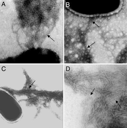
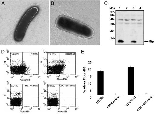
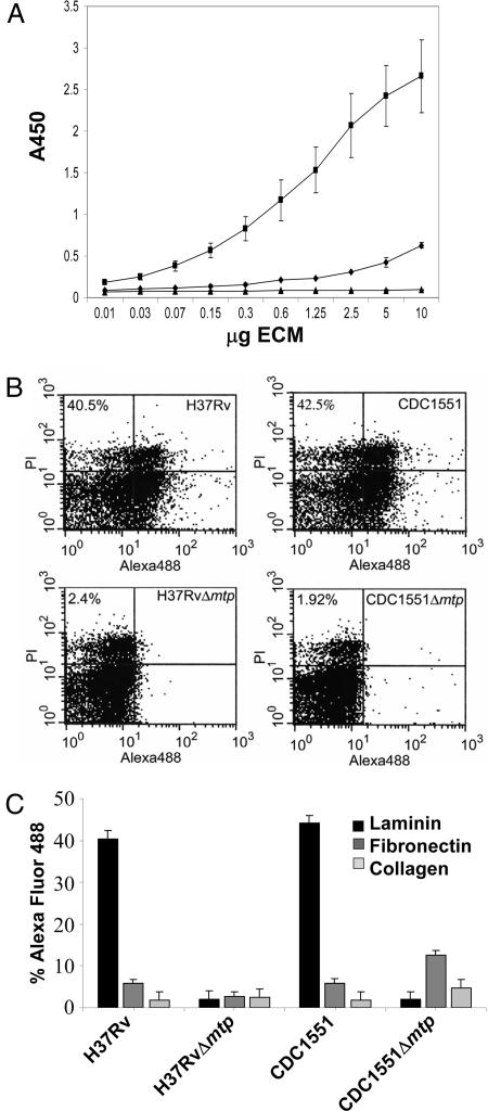
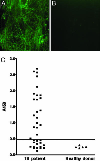
Mycobacterium tuberculosis is responsible for nearly 3 million human deaths worldwide every year. Understanding the mechanisms and bacterial factors responsible for the ability of M. tuberculosis to cause disease in humans is critical for the development of improved treatment strategies. Many bacterial pathogens use pili as adherence factors to colonize the host. We discovered that M. tuberculosis produces fine (2- to 3-nm-wide), aggregative, flexible pili that are recognized by IgG antibodies contained in sera obtained from patients with active tuberculosis, indicating that the bacilli produce pili or pili-associated antigen during human infection. Purified M. tuberculosis pili (MTP) are composed of low-molecular-weight protein subunits encoded by the predicted M. tuberculosis H37Rv ORF, designated Rv3312A. MTP bind to the extracellular matrix protein laminin in vitro, suggesting that MTP possess adhesive properties. Isogenic mtp mutants lost the ability to produce Mtp in vitro and demonstrated decreased laminin-binding capabilities. MTP shares morphological, biochemical, and functional properties attributed to bacterial pili, especially with curli amyloid fibers. Thus, we propose that MTP are previously unidentified host-colonization factors of M. tuberculosis.


Conflict of interest statement

The authors declare no conflict of interest.

Figures

Fig. 1.
Fig. 1.
Demonstration of pili production by M. tuberculosis strains. Transmission electron micrographs of negatively stained M. tuberculosis strains H37Rv (A), avirulent H37Ra (B), clinical isolate CDC1551 (C), and purified MTP (D). Black arrows point to MTP pili fibers. (Magnification: ×45,000.)
Fig. 2.
Fig. 2.
Isogenic M. tuberculosis mtp mutants lack production of pili fibers. (A and B) Electron micrographs of H37RvΔ mtp (A) and CDC1551Δ mtp (B) showing no filamentous structures. (Magnification: ×25,000.) (C) Anti-Mtp Western blot of whole bacteria extracts showing production of the pilus protein in H37Rv (lane 1) and CDC1551 (lane 3) and absence of the pilin in both H37RvΔ mtp (lane 2) and CDC1551Δ mtp (lane 4). (D) Surface detection of MTP in wild-type parental and derivative mtp deletion strains by flow cytometry (n = 50,000) using anti-Mtp antisera. MTP–antibody complexes were detected by using Alexa Fluor conjugate. (E) Plot of the flow cytometry values obtained in D.
Fig. 3.
Fig. 3.
MTP binds to laminin. (A) ELISA-based assay to measure the binding of increasing concentrations of fibronectin (♦), laminin (■), and collagen type IV (▴) to MTP-coated plates. Binding was detected by using either rabbit anti-fibronectin, anti-laminin, or mouse anti-collagen type IV. All ELISA values given are averaged from triplicate experiments. Error bars represent the standard deviation from the mean. (B) Flow cytometry demonstration of binding of laminin to MTP-producing M. tuberculosis but not to mtp mutants. Binding to fibronectin and collagen was negligible (n = 50,000). Binding was determined by using anti-ECM antibodies described above followed by detection using Alexa Fluor conjugate. (C) Bars indicate relative Alexa Fluor signal from flow cytometry. Data are presented from triplicate experiments.
Fig. 4.
Fig. 4.
MTP are produced during human TB infection. (A) IF showing that MTP incubated with TB patient sera produced fluorescent MTP fibers. (B) No reaction was observed when MTP was incubated with sera from healthy donors. (Original magnification: ×1,000.) (C) Sera from TB patients (n = 36) and from healthy donors (n = 5) were tested for anti-MTP IgG antibodies by ELISA. Most of the patients' sera (60%) showed a significant IgG titer against immobilized MTP fibers. Results obtained at sera diluted 1:3,200 are shown. The horizontal line represents the cut-off value of two times the average ELISA reading of healthy control sera. All of the sera positive by ELISA produced a positive IF reaction as shown in A.

References

    1. Dye C, Scheele S, Dolin P, Pathania V, Raviglione MC. J Am Med Assoc. 1999;282:677–686. - PubMed
    1. Frieden TR, Sterling TR, Munsiff SS, Watt CJ, Dye C. Lancet. 2003;362:887–899. - PubMed
    1. Armstrong JA. J Exp Med. 1971;134:713–740. - PMC - PubMed
    1. Shepard CC. Proc Soc Exp Biol Med. 1955;90:392–396. - PubMed
    1. Schlesinger LS, Bellinger-Kawahara CG, Payne NR, Horwitz MA. J Immunol. 1990;144:2771–2780. - PubMed

Publication types

MeSH terms