. 2007 Mar 6;104(10):3765-70.
doi: 10.1073/pnas.0609211104. Epub 2007 Feb 22.

Regulation of poly(ADP-ribose) polymerase 1 activity by the phosphorylation state of the nuclear NAD biosynthetic enzyme NMN adenylyl transferase 1

Affiliations

Regulation of poly(ADP-ribose) polymerase 1 activity by the phosphorylation state of the nuclear NAD biosynthetic enzyme NMN adenylyl transferase 1

Felicitas Berger et al. Proc Natl Acad Sci U S A. .

Abstract

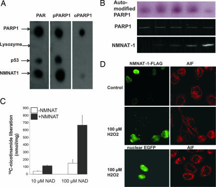
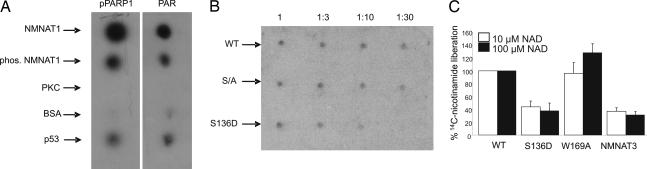
Nuclear NAD(+) metabolism constitutes a major component of signaling pathways. It includes NAD(+)-dependent protein deacetylation by members of the Sir2 family and protein modification by poly(ADP-ribose) polymerase 1 (PARP-1). PARP-1 has emerged as an important mediator of processes involving DNA rearrangements. High-affinity binding to breaks in DNA activates PARP-1, which attaches poly(ADP-ribose) (PAR) to target proteins. NMN adenylyl transferases (NMNATs) catalyze the final step of NAD(+) biosynthesis. We report here that the nuclear isoform NMNAT-1 stimulates PARP-1 activity and binds to PAR. Its overexpression in HeLa cells promotes the relocation of apoptosis-inducing factor from the mitochondria to the nucleus, a process known to depend on poly(ADP-ribosyl)ation. Moreover, NMNAT-1 is subject to phosphorylation by protein kinase C, resulting in reduced binding to PAR. Mimicking phosphorylation, substitution of the target serine residue by aspartate precludes PAR binding and stimulation of PARP-1. We conclude that, depending on its state of phosphorylation, NMNAT-1 binds to activated, automodifying PARP-1 and thereby amplifies poly(ADP-ribosyl)ation.


Conflict of interest statement

The authors declare no conflict of interest.

Figures

Fig. 1.
Fig. 1.
PKC-mediated phosphorylation of NMNAT-1. (A) Recombinant NMNAT-1 (500 ng) was incubated with PKC or casein kinase II (CKII) (10 units) and [γ-32P]ATP. Proteins were then separated by SDS/PAGE. The autoradiograph of the gel is shown. (B) NMNAT-1 (500 ng) was incubated with nuclear extracts (1 μg) in the presence of [γ-32P]ATP and phorbol myristate acetate (PMA) or BIM as indicated. Proteins were separated by SDS/PAGE, and gels were analyzed by autoradiography. (C) Human fibroblasts were incubated with [32P]orthophosphate for 8 h. Where indicated, BIM (1 μM) was added 1 h before cell lysis. After immunoprecipitation of endogenous NMNAT-1, proteins were separated by SDS/PAGE. Labeled proteins were visualized by autoradiography. The numbers on the left indicate the mobility of marker proteins. The asterisk shows the position of NMNAT-1.
Fig. 2.
Fig. 2.
Identification of the NMNAT-1 phosphorylation site as serine 136. (A) Recombinant NMNAT-1 (25 μg) was 32P-phosphorylated by PKC and then subjected to tryptic digestion. The generated peptides were separated by HPLC. The elution profile is shown. The upper, black trace shows absorbance at 214 nm. The lower, gray trace shows radioactivity of the collected fractions. AUFS, Absorbance units full scale. (B) The fraction containing a radioactive peptide was analyzed by MALDI-TOF mass spectrometry. The relevant part of the mass spectrum is presented. (C) The obtained peptide masses and the corresponding stretches of the NMNAT-1 sequence are shown. (D) WT NMNAT-1 or the S136A mutant was incubated with PKC or HeLa nuclear extracts as indicated in the presence of [γ-32P]ATP. Proteins were separated by SDS/PAGE (PKC Left and Nuclear Extracts Left) and then subjected to autoradiography (PKC Right and Nuclear Extracts Right).
Fig. 3.
Fig. 3.
Characterization of generated NMNAT-1 proteins. FLAG-tagged WT and mutant proteins were overexpressed in HEK 293 cells. The FLAG tag and nuclei were visualized by fluorescence microscopy using FLAG-specific antibodies and DAPI, respectively (as indicated). Western blots of the purified NMNAT-1 proteins overexpressed in E. coli (as His6-tagged proteins) were probed with antibodies specific for the His6 tag or against WT NMNAT-1 (as indicated). The catalytic activities of the purified recombinant proteins are given as the average value obtained from two independent protein preparations. The activity of the W169A mutant, if any, was below the detection level.
Fig. 4.
Fig. 4.
NMNAT-1 associates with PAR and automodified PARP-1 and accelerates poly(ADP-ribosyl)ation and AIF translocation. (A) Specific interaction of NMNAT-1 with free or PARP-1-bound PAR was visualized by spotting the indicated proteins (0.5 μg) on a nitrocellulose membrane. The membrane was then incubated with 32P-labeled oligo(ADP-ribosyl)ated PARP-1 (oPARP-1, 0.25 μg), poly(ADP-ribosyl)ated PARP-1 (pPARP-1, 0.25 μg), or protein-free PAR (PAR, 5 μM). After washing, specific binding was visualized by autoradiography. (B) PARP-1 (25 ng) was incubated with 32P-NAD+ in the absence or presence of NMNAT-1 in a mass ratio (PARP-1/NMNAT-1) of 1:2, 1:4, 1:10, or 1:20. Thereafter, the proteins were separated by SDS/PAGE. (Top) The autoradiograph of the gel is shown. (Middle and Bottom) Proteins were visualized by the fluorescent SYPRO Ruby stain (Invitrogen, Carlsbad, CA). (C) PARP-1 (100 ng) was incubated with nicotinamide/[14C]NAD+ in the presence or absence of NMNAT-1 (mass ratio of 1:5). [14C]Nicotinamide released during automodification of PARP-1 was identified by HPLC using radioisotope detection. (D) PAR-mediated AIF translocation is enhanced by NMNAT-1 overexpression. FLAG-tagged NMNAT-1 was transiently overexpressed in HeLa cells. (Bottom Left) As a control, nuclear EGFP was overexpressed. The cells were treated with 100 μM H2O2 as indicated. FLAG-tagged NMNAT-1 and AIF were visualized by immunostaining.
Fig. 5.
Fig. 5.
Phosphorylated NMNAT-1 has less affinity for automodified PARP-1 and does not stimulate PARP-1 activity. (A) Recombinant NMNAT-1 was phosphorylated by PKC, as indicated. Proteins (0.5 μg) were spotted onto nitrocellulose. The membrane was then incubated with 32P-labeled poly(ADP-ribosyl)ated PARP-1 or free polymers. Thereafter, the membrane was washed, and specific binding was visualized by autoradiography. (B) Serial dilutions of the indicated recombinant NMNAT-1 proteins were spotted onto nitrocellulose. The membrane was then incubated with 32P-labeled poly(ADP-ribosyl)ated PARP-1 (0.25 μg/ml), washed, and subjected to autoradiography. S/A, serial mutant in which serines 135 and 136 were replaced by alanine residues. (C) PARP-1 was incubated with [14C]nicotinamide/NAD+ in the presence of the indicated NMNAT-1 proteins or NMNAT-3, as a control. [14C]Nicotinamide released during automodification of PARP-1 was analyzed by HPLC using radioisotope detection.

References

    1. Magni G, Amici A, Emanuelli M, Raffaelli N, Ruggieri S. Adv Enzymol Relat Areas Mol Biol. 1999;73:135–182. - PubMed
    1. Berger F, Lau C, Dahlmann M, Ziegler M. J Biol Chem. 2005;280:36334–36341. - PubMed
    1. Schweiger M, Hennig K, Lerner F, Niere M, Hirsch-Kauffmann M, Specht T, Weise C, Oei SL, Ziegler M. FEBS Lett. 2001;492:95–100. - PubMed
    1. Anderson RM, Bitterman KJ, Wood JG, Medvedik O, Cohen H, Lin SS, Manchester JK, Gordon JI, Sinclair DA. J Biol Chem. 2002;277:18881–18890. - PubMed
    1. Berger F, Ramirez-Hernandez MH, Ziegler M. Trends Biochem Sci. 2004;29:111–118. - PubMed

Publication types