. 2007 Mar 6;104(10):3829-34.
doi: 10.1073/pnas.0610055104. Epub 2007 Feb 26.

c-Myb is required for progenitor cell homeostasis in colonic crypts

Affiliations

c-Myb is required for progenitor cell homeostasis in colonic crypts

Jordane Malaterre et al. Proc Natl Acad Sci U S A. .

Abstract

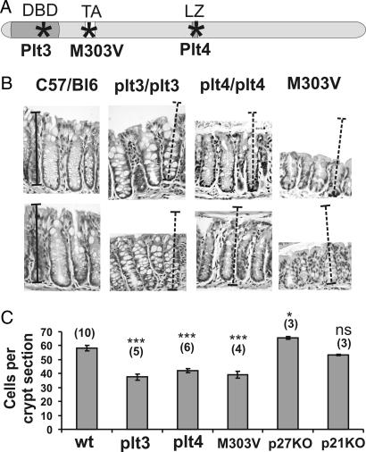
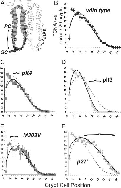
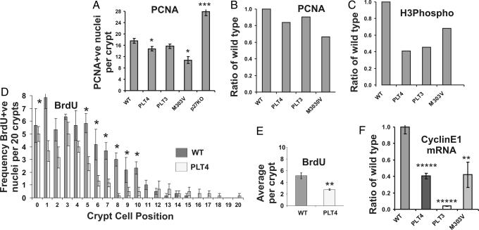
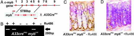
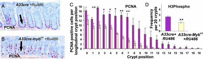
The colonic crypt is the functional unit of the colon mucosa with a central role in ion and water reabsorption. Under steady-state conditions, the distal colonic crypt harbors a single stem cell at its base that gives rise to highly proliferative progenitor cells that differentiate into columnar, goblet, and endocrine cells. The role of c-Myb in crypt homeostasis has not been elucidated. Here we have studied three genetically distinct hypomorphic c-myb mutant mouse strains, all of which show reduced colonic crypt size. The mutations target the key domains of the transcription factor: the DNA binding, transactivation, and negative regulatory domains. In vivo proliferation and cell cycle marker studies suggest that these mice have a progenitor cell proliferation defect mediated in part by reduced Cyclin E1 expression. To independently assess the extent to which c-myb is required for colonic crypt homeostasis we also generated a novel tissue-specific mouse model to allow the deletion of c-myb in adult colon, and using these mice we show that c-Myb is required for crypt integrity, normal differentiation, and steady-state proliferation.


Conflict of interest statement

The authors declare no conflict of interest.

Figures

Fig. 1.
Fig. 1.
Hypomorphic c-myb mutant mice have shorter crypts than wild-type mice. (A) Murine c-Myb diagram marking (∗) the location of amino acid substitutions within the DNA binding domain (Plt3), the leucine zipper region (Plt4) (8), and the transactivation domain (M303V) (9). (B) H&E-stained sections of wild-type, plt3/plt3, plt4/plt4, and M303V/M303V distal colons show that the mutant crypts are shorter than wild type. Relative length size bars are shown layered over normal distal colonic crypts, and these have been transferred to panels representing the three hypomorphs. (C) Morphometric analysis of the sections indicates that the hypomorphic mutant crypts are significantly shorter than wild-type C57BL/6 crypts whereas p27−/− crypts are significantly longer. p21−/− crypts are indistinguishable from wild type. Bars represent mean ± SEM (number of mice analyzed). ∗∗∗, P > 0.0001 (ANOVA).
Fig. 2.
Fig. 2.
Hypomorphic mutant crypts show reduced proliferation. (A) Model of a distal colonic crypt showing the presumed location of the stem cell (SC) and region of proliferation ascribed to the progenitor cell population (PC). The crypt cell position referenced to position zero represents the location of the presumptive stem cells. (B–F) When crypts were stained for proliferation marker PCNA, the extent and distribution of antigen-positive cells was significantly different in the plt3/plt3, plt4/plt4, M03V/M303V, and p27 knockout mice. Horizontal brackets demark where there are significant differences from wild-type crypts. c-myb hypomorphs show significantly less proliferation in these regions. The data also show that proliferation in the p27−/− crypts is more extensive than wild-type crypts, consistent with the longer crypts evident in these mice. Quadratic trend lines are shown to allow visual comparisons between the mutant and wild-type (hashed lined) crypts.
Fig. 3.
Fig. 3.
Proliferation markers indicate that hypomorphic c-myb mice have a defect in cell cycle progression. (A) Collectively, the total number of PCNA-positive cells per crypt for each strain of mice was significantly different for all mutants with the exception of PLT3 mutants. (B) To allow a further assessment of a potential defect in cell cycle progression each data set was analyzed as a ratio to wild type to show the reduced PCNA staining in mutants. (C) This type of analysis was extended to include phospho-histone-3 expression whereby it would appear that the ratios of this marker of G2/M phase cells are less than those observed for cycling PCNA-stained cells. (D and E) PLT4 mutant and wild-type mice were further assessed for the extent of BrdU staining on the basis of crypt position (D) and total per crypt (E). Both analyses suggested significantly less S-phase progression in PLT4 mutant mice compared with wild-type littermates. Bars represent mean ± SEM. ∗∗, P < 0.01; ∗, P < 0.05 (ANOVA). (F) Real-time RT-PCR studies on colonic crypt RNA isolated from wild-type (n = 6), PLT3 (n = 3), PLT4 (n = 3), and M303V (n = 3) mice show that Cyclin E1 expression is lower in these hypomorphic mutant mice, perhaps explaining the defect in cell cycle progression particularly at the crypt base.
Fig. 4.
Fig. 4.
Tissue-specific conditional deletion of c-myb in distal colon leads to a reduction of proliferation in colonic crypts. (A) Schematic of the strategy for the conditional deletion of the c-myb locus (14) whereby exons 3–6 are excised after the activation of A33CrePR2 activity by RU486. (B) An image of an agarose gel with ethidium bromide-stained PCR products generated by using primers (marked by red arrows in A) indicated successful deletion of exons 3–6 in the presence of RU486 (+). Costaining of colonic crypts for c-Myb (C) and PAS (D) shows that the intensity of PAS staining (as an indicator of goblet cell differentiation) increases with the loss of c-Myb.
Fig. 5.
Fig. 5.
c-myb is required for proliferation in colonic crypts. (A and B) Colon sections were stained for PCNA in nonrecombined colonic crypts (A) and in recombined crypts where reduced proliferation is evident (B). (C) Quantitation of PCNA staining in discernable crypts in nonrecombined and RU486-treated mice is shown for each crypt cell position, indicating that after Cre activation with RU486 the proportion of cells per crypt is significantly reduced. (D) Crypts stained for phospho-histone-3 to examine cell cycle progression into the G2/M phases of cell cycle indicate that the crypts with deleted c-myb had fewer cells in the G2/M phase of the cell cycle compared with unrecombined crypts. Bars represent mean ± SEM (n = 3 per treatment). ∗∗, P < 0.01; ∗, P < 0.05 (ANOVA).

References

    1. Potten CS. Philos Trans R Soc London B. 1998;353:821–830. - PMC - PubMed
    1. Gavrieli Y, Sherman Y, Ben-Sasson SA. J Cell Biol. 1992;119:493–501. - PMC - PubMed
    1. Gordon JI, Hermiston ML. Curr Opin Cell Biol. 1994;6:795–803. - PubMed
    1. Rosenthal MA, Thompson MA, Ellis S, Whitehead RH, Ramsay RG. Cell Growth Differ. 1996;7:961–967. - PubMed
    1. Mucenski ML, McLain K, Kier AB, Swerdlow SH, Schreiner CM, Miller TA, Pietryga DW, Scott WJ, Jr, Potter SS. Cell. 1991;65:677–689. - PubMed

Publication types