. 2007 Mar 6;104(10):3871-6.
doi: 10.1073/pnas.0611637104. Epub 2007 Feb 28.

Interaction with the mammary microenvironment redirects spermatogenic cell fate in vivo

Affiliations

Interaction with the mammary microenvironment redirects spermatogenic cell fate in vivo

Corinne A Boulanger et al. Proc Natl Acad Sci U S A. .

Abstract

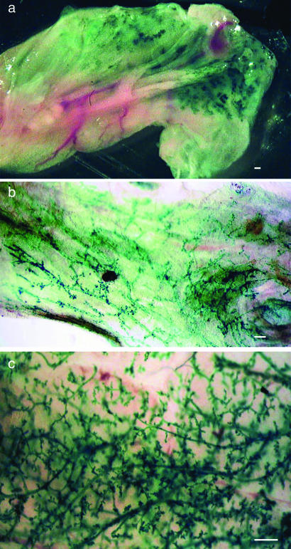
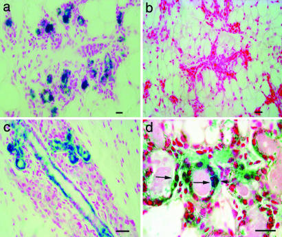
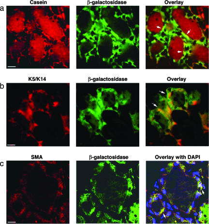
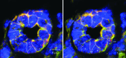
Previously, we characterized a parity-induced mammary epithelial cell population that possessed the properties of pluripotency and self-renewal upon transplantation. These cells were lineally marked by the expression of beta-galactosidase (LacZ) as a result of mammary-specific activation of a reporter gene through Cre-lox recombination during pregnancy. We used this experimental model to determine whether testicular cells would alter their cell fate upon interaction with the mammary gland microenvironment during pregnancy, lactation, and involution. Adult testicular cells, isolated from seminiferous tubules, were mixed with limiting dilutions of dispersed mammary epithelial cells and injected into epithelium-divested mammary fat pads. The host mice were bred 6-8 weeks later and examined 20-30 days postinvolution. This approach allowed for the growth of mammary tissue from the injected cells and transient activation of the whey acidic protein promoter-Cre gene during pregnancy and lactation, leading to Cre-lox recombination and constitutive expression of LacZ from its promoter. Here we show that cells from adult seminiferous tubules interact with mammary epithelial cells during regeneration of the gland. They adopt mammary epithelial progenitor cell properties, including self-renewal and the production of cell progeny, which differentiate into functional mammary epithelial cells. Our results provide evidence for the ascendancy of the tissue microenvironment over the intrinsic nature of cells from an alternative adult tissue.


Conflict of interest statement

The authors declare no conflict of interest.

Figures

Fig. 1.
Fig. 1.
Detection of male-derived cells in chimeric outgrowths. (a) X-Gal-stained whole mammary fat pad from postpregnant host mouse reveals numerous LacZ+ cells within the gland. (b) After removal of fat, LacZ cells were found uniformly distributed along the mammary ducts in this whole mount. (c) LacZ+ cells in a whole mount from a second-generation transplant showing the self-renewal of male progeny. (Scale bars: 1.0 mm.)
Fig. 2.
Fig. 2.
Tissue section analysis of X-Gal-stained outgrowths. (a) Cross-section through mammary ducts showing the distribution of LacZ+ epithelial cells among negative stroma. (b) Section from the host mammary gland from same mouse. (c) Longitudinal section of duct in chimeric outgrowth with LacZ+ epithelium. (d) Higher magnification of LacZ+ epithelial cells in secretory acinus of pregnant host showing blue luminal cells (long arrows) adjacent to blue myoepithelial cell (short arrow). (Scale bars: 20 μm.)
Fig. 3.
Fig. 3.
Male cell progeny may express luminal or myoepithelial cell markers. (a) Colocalization of casein and β-galactosidase in chimeric outgrowths of lactating murine mammary glands. Arrows indicate luminal cells that express both casein and β-galactosidase. (b and c) Male cells expressing β-galactosidase and keratin 5 (K5)/keratin 14 (K14) (b) or smooth muscle actin (SMA) (c), indicating a myoepithelial fate. Arrows in b indicate cells expressing both K5/K14 and β-galactosidase; arrows in c indicate cells expressing both β-galactosidase and smooth muscle actin. (Scale bars: a and b, 20 μm; c, 10 μm.)
Fig. 4.
Fig. 4.
X and Y chromosome FISH analysis was performed on paraffin-embedded, 6-μm sections of mammary glands containing chimeric outgrowths. A mammary secretory structure is shown with the X chromosome labeled in red (x), the Y chromosome labeled in green (y), and the nuclei stained with DAPI. Serial 1.0-μm slices photographed under three-color confocal microscopy reveal the presence of male cells juxtaposed with female cells in the same acinus. (Scale bars: 5.0 μm.)

References

    1. Li L, Xie T. Annu Rev Cell Dev Biol. 2005;21:605–631. - PubMed
    1. Deome KB, Faulkin LJ, Jr, Bern HA, Blair PB. Cancer Res. 1959;19:515–520. - PubMed
    1. Daniel C, DeOme K, Young L, Blair P, Faulkin L. Proc Natl Acad Sci USA. 1968;61:53–60. - PMC - PubMed
    1. Daniel CW, Young LJ. Exp Cell Res. 1971;65:27–32. - PubMed
    1. Young LJ, Medina D, DeOme KB, Daniel CW. Exp Gerontol. 1971;6:49–56. - PubMed

LinkOut - more resources