. 2007 Mar 6;104(10):3877-82.
doi: 10.1073/pnas.0700416104. Epub 2007 Feb 26.

Mechanisms controlling the acquisition of a cardiac phenotype by liver stem cells

Affiliations

Mechanisms controlling the acquisition of a cardiac phenotype by liver stem cells

Barbara J Muller-Borer et al. Proc Natl Acad Sci U S A. .

Abstract

The mechanisms underlying stem cell acquisition of a cardiac phenotype are unresolved. We studied early events during the acquisition of a cardiac phenotype by a cloned adult liver stem cell line (WB F344) in a cardiac microenvironment. WB F344 cells express a priori the transcription factors GATA4 and SRF, connexin 43 in the cell membrane, and myoinositol 1,4,5-triphosphate receptor in the perinuclear region. Functional cell-cell communication developed between WB F344 cells and adjacent cocultured cardiomyocytes in 24 h. De novo cytoplasmic [Ca(2+)](c) and nuclear [Ca(2+)](nu) oscillations appeared in WB F344 cells, synchronous with [Ca(2+)](i) transients in adjacent cardiomyocytes. The [Ca(2+)] oscillations in the WB F344 cells, but not those in the cardiomyocytes, were eliminated by a gap junction uncoupler and reappeared with its removal. By 24 h, WB F344 cells began expressing the cardiac transcription factors Nkx2.5, Tbx5, and cofactor myocardin; cardiac proteins 24 h later; and a sarcomeric pattern 4-6 days later. Myoinositol 1,4,5-triphosphate receptor inhibition suppressed WB F344 cell [Ca(2+)](nu) oscillations but not [Ca(2+)](c) oscillations, and L-type calcium channel inhibition eliminated [Ca(2+)] oscillations in cardiomyocytes and WB F344 cells. The use of these inhibitors was associated with a decrease in Nkx2.5, Tbx5, and myocardin expression in the WB F344 cells. Our findings suggest that signals from cardiomyocytes diffuse through shared channels, inducing [Ca(2+)] oscillations in the WB F344 cells. We hypothesize that the WB F344 cell [Ca(2+)](nu) oscillations activate the expression of a cardiac specifying gene program, ushering in a cardiac phenotype.


Conflict of interest statement

The authors declare no conflict of interest.

Figures

Fig. 1.
Fig. 1.
Gap junction-mediated cell–cell communication recorded in early (24–48 h) WB F344/cardiomyocyte cocultures. (A and B) Calcein-labeled coculture with WB F344 cell 1 and cell 2 adjacent to cardiomyocytes before laser excitation (A) and immediately after exposure to high-intensity laser excitation (B). (C) Fluorescence recovery in WB F344 cell 1 through shared gap junction channels with adjacent cardiomyocytes. It is presumed that WB F344 cell 2 was not functionally coupled with adjacent myocytes because no fluorescence recovery was recorded. (D) Graph of fluorescence recovery vs. time in WB F344 cell 1 and cell 2.
Fig. 2.
Fig. 2.
Calcium signals acquired from undifferentiated WB F344 cells and adjacent cardiomyocytes. (Upper) Image of dsRed WB F344 cell adjacent to cardiomyocyte in coculture labeled with fluo-4 AM. Confocal line scan microscopy was used to record intracellular calcium oscillations along the dotted line. (Lower) Tracings correspond to the areas marked by the color-coded dots in the line scan. Shown are signal-averaged calcium signals at the immediate cardiomyocyte (green)/WB F344 cell (red) interface, 4 μm away from the cell interface in the WB F344 cell (blue), and 4 μm away from the interface in the cardiomyocyte (black). Decreased amplitude of the [Ca2+] signal is demonstrated in the WB F344 cell with distance from the cell interface (red and blue lines) suggesting a diffusion-mediated signal but not in the cardiomyocyte (green and black lines), where a uniform [Ca2+]i within the cardiomyocyte is believed to be mediated by a calcium-induced calcium release process. The calcium oscillations in the WB F344 cells depend on direct contact between the WB F344 cells and adjacent myocytes. WB F344 cells cultured alone or distant from cardiomyocytes did not demonstrate oscillations even when paced, suggesting that a humoral factor secreted into the medium did not induce these oscillations. (Scale bar: 10 μm.)
Fig. 3.
Fig. 3.
Inhibition of calcium signals acquired from undifferentiated WB F344 cells cocultured with cardiomyocytes. (A) Representative image of undifferentiated WB F344 cell adjacent to cardiomyocyte in coculture. Cardiomyocyte [Ca2+]i transients and adjacent WB F344 cell [Ca2+]c oscillations or cardiomyocyte [Ca2+]i transients and adjacent WB F344 cell [Ca2+]n oscillations were acquired by line scan confocal microscopy in the region along dotted line 1 or line 2, respectively. (B) WB F344 cell [Ca2+]c oscillations (red) were synchronous with the cardiomyocyte [Ca2+]i transients (green) during control conditions. The line scan was recorded from an area that corresponds to the dotted line 1 in A. WB F344 cell [Ca2+]c oscillations were abolished in the presence of the gap junction uncoupler carbenoxolone, whereas the cardiomyocyte [Ca2+]i transients were maintained. Approximately 30 min after the carbenoxolone was washed out of the culture medium, the WB F344 cell [Ca2+]c oscillations were restored. (C) Inhibition of WB F344 cell [Ca2+]nu oscillations. Cytoplasmic [Ca2+]c (red) and nuclear [Ca2+]nu (black) signals were recorded in a WB F344 cell in coculture before and after the addition of the IP3R inhibitor 2-APB. The tracings represent calcium signals recorded from a WB F344 cell nuclear region that corresponds to dotted line 2 in A. The WB F344 cell [Ca2+]nu signals (black) were significantly suppressed in the presence of the IP3R inhibitor. (D) IP3R was localized by immunocytochemistry to the perinuclear region (white arrow and green fluorescence in D) and the endoplasmic reticulum (red arrows) of WB F344 cells. D is a merged image of FITC fluorescence and DAPI nuclear counterstain (×63 oil immersion objective lens).
Fig. 4.
Fig. 4.
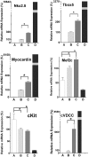
RNA levels (expressed as fold change) of cardiac transcription factors, Nkx2.5, Tbx5, myocardin, and Mef2c, WB F344 cells cultured alone (column A), WB F344 cells cocultured with heart cells for 18–48 h (column B), WB F344 cells cocultured with heart cells for 48–72 h (column C), and cardiac cells cultured alone (column D). Gene reprogramming is noted by the decrease in expression of c-Kit and the increase in expression of the cVDCC with time in culture in the same conditions. The bars show mean ± SEM. ∗, P < 0.001. (See primer sequences in SI Table 1.)
Fig. 5.
Fig. 5.
Suppression of cardiac transcription factor expression with inhibitors. Expression (noted in fold change) of the cardiac transcription factors Nkx2.5, Tbx5, and myocardin in WB F344 cells cultured alone (column A), WB F344 cells cocultured with heart cells for 18–24 h (column B), WB F344 cells cocultured with heart cells in the presence of the IP3R inhibitor 2-APB for 18–24 h (column C), and WB F344 cells cocultured with heart cells in the presence of the cVDCC inhibitor nifedipine for 18–24 h (column D). The bars show mean ± SEM. ∗, P < 0.001.
Fig. 6.
Fig. 6.
Polymorphic microsatellite markers. By using the D9U1A7 set of microsatellite markers, the amplicons from the control or FACS-harvested Fischer WB F344 cells DNA (lanes 4 and 5) are shifted down compared with the PCR product from control Sprague–Dawley DNA (lane 3, white asterisks in Top). Lane 1, DNA ladder; lane 2, no DNA control. By using a second set of markers, D17U1A2, three amplicons (white asterisks in Middle) are present only in the Sprague–Dawley control DNA (lane 3) but not in the harvested WB F344 cells. By using a third set of markers, D1U1A14, the amplicons from the Fischer WB F344 DNA (lanes 4 and 5) are shifted up compared with the PCR product from the Sprague–Dawley DNA (white asterisks in Bottom). (See primer sequences in SI Table 2.)

References

    1. Anversa P, Leri A, Kajstura J. J Am Coll Cardiol. 2006;47:1769–1776. - PubMed
    1. Murry CE, Reinecke H, Pabon LM. J Am Coll Cardiol. 2006;47:1777–1785. - PubMed
    1. Alvarez-Dolado M, Pardal R, Garcia-Verdugo JM, Fike JR, Lee HO, Pfeffer K, Lois C, Morrison SJ, Alvarez-Buylla A. Nature. 2003;425:968–973. - PubMed
    1. Muller-Borer BJ, Cascio WE, Anderson PA, Snowwaert JN, Frye JR, Desai N, Esch GL, Brackham JA, Bagnell CR, Coleman WB, et al. Am J Pathol. 2004;165:135–145. - PMC - PubMed
    1. Kajstura J, Rota M, Whang B, Cascapera S, Hosoda T, Bearzi C, Nurzynska D, Kasahara H, Zias E, Bonafe M, et al. Circ Res. 2005;96:127–137. - PubMed

Publication types