. 2007 Mar 6;104(10):3949-54.
doi: 10.1073/pnas.0609783104. Epub 2007 Feb 27.

Targeted deletion of Wwox reveals a tumor suppressor function

Affiliations

Targeted deletion of Wwox reveals a tumor suppressor function

Rami I Aqeilan et al. Proc Natl Acad Sci U S A. .

Abstract

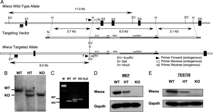
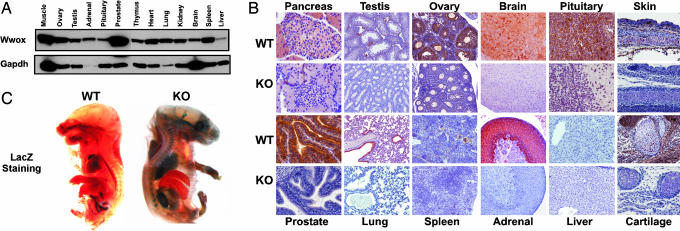
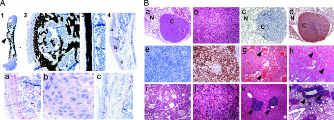
The WW domain-containing oxidoreductase (WWOX) spans the second most common fragile site of the human genome, FRA16D, located at 16q23, and its expression is altered in several types of human cancer. We have previously shown that restoration of WWOX expression in cancer cells suppresses tumorigenicity. To investigate WWOX tumor suppressor function in vivo, we generated mice carrying a targeted deletion of the Wwox gene and monitored incidence of tumor formation. Osteosarcomas in juvenile Wwox(-/-) and lung papillary carcinoma in adult Wwox(+/-) mice occurred spontaneously. In addition, Wwox(+/-) mice develop significantly more ethyl nitrosourea-induced lung tumors and lymphomas in comparison to wild-type littermate mice. Intriguingly, these tumors still express Wwox protein, suggesting haploinsuffiency of WWOX itself is cancer predisposing. These results indicate that WWOX is a bona fide tumor suppressor.


Conflict of interest statement

The authors declare no conflict of interest.

Figures

Fig. 1.
Fig. 1.
Targeted disruption of the Wwox gene. (A) The Wwox genomic locus was altered by using a targeting vector that replaced ≈6 kb of genomic sequences that include exons 2, 3, and 4 (vertical dark boxes, E2, E3, E4) with a targeting cassette containing a chimeric sequence derived from the in-frame fusion of LacZ and Neo genes. A 5′ genomic probe (horizontal dark box) that recognized an ≈11-kb WT fragment and an ≈8-kb targeted EcoRV fragment was used for genotyping by Southern blotting. Three primers (arrows), a shared 5′ primer (F), distinct 3′ primers that recognized WT locus (R), and the other 3′ primers specific for LacZ-Neo sequence (R1) allowed PCR genotyping. (B) Southern blot analysis of genomic DNA extracted from mouse tails showing WT, HT, and homozygous (KO) mutant genotypes. (C) PCR genotype of DNA extracted from mouse tails showing the different genotypes. B6129F1-F4 mice were genotyped at age 14–21 days. M, marker. (D and E) Western blot analysis of mouse embryonic fibroblasts and testis with monoclonal anti-WWOX antibody indicating undetectable levels of Wwox in homozygous mutant mice. GAPDH levels were used for normalization.
Fig. 2.
Fig. 2.
Wwox expression pattern in mouse tissue. (A) Protein lysate from the indicated tissues was probed with monoclonal anti-WWOX antibody or GAPDH antibody as control. (B) Immunohistochemical staining of Wwox in main tissues from WT and KO mice with polyclonal anti-WWOX antibody, indicating high expression of Wwox in different epithelial tissues and in the endocrine system. Stained sections from pancreases, testis, ovary, pituitary, brain (brainstem), skin, prostate, lung, spleen, adrenal gland (cortex), liver, and cartilage primordium of the skull are shown. (C) LacZ staining of embryo from E17.5. E17.5 embryos were fixed in paraformaldehyde, stained with X-Gal, and analyzed by dissecting light microscope for gross morphology and LacZ expression (blue). Homozygous mutant embryo shows expression of LacZ in brain, lung, kidney, adrenal gland, stomach, and bones, whereas WT embryo is not stained.
Fig. 3.
Fig. 3.
Tumor phenotype in Wwox mutant mice. (A) Formation of osteosarcomas in Wwox null mice. Typical histological appearance is shown for one tumor (A1–A3, toluidine blue with von Kossa stain). Calcified chondroid tissue forms the diaphysis (A1, arrowhead) with immature trabeculae in the osteosarcoma (A2). A section of diaphysis of WT mouse from the same litter showing a single layer of osteoblasts constituting the endosteal and periosteal surfaces of cortical bone (A3). Decalcified femur bone, age 17 days (A4), reveals a cartilage cap under the periosteum with enlarged hypertrophic cells forming a growth plate (Aa). Proliferating cells adjacent to this lesion appear to be transformed (Ab). (Ac) Hyperproliferative periosteal cells along the diaphysis. (B) Histopathology of tumors in Wwox+/− mice. (Ba) Lung (H&E staining) nodule composed of epithelial proliferation with glandular and papillary growth pattern (bronchiolo-alveolar carcinoma). N (normal) versus C (carcinoma). (Magnification: × 200.) (Bb) Higher magnification (H&E staining) of the lung nodule shown in Ba, indicating marked nuclear and cellular atypia with rare atypical mitoses. (Magnification: ×400.) (Bc) Immunohistochemistry for Ki67 showing low proliferation rate in tumor cells in a lung nodule. (Magnification: ×200.) (Bd) Immunohistochemistry for WWOX showing positive expression in tumor cells in a lung nodule. (Magnification: ×100.) (Be) Spleen (H&E staining) monomorphic lymphopid population expanding the red pulp: diffuse large B cell lymphoma. (Magnification: ×400.) (Bf) Immunohistochemistry for Ki67 showing intense positivity in the lymphoblastic population in spleen. (Magnification: ×100.) (Bg) Hemangioma in liver (H&E staining). Arrows show large vascular channels. (Magnification: ×100.) (Bh) Large mass of malignant chondroblasts surrounded by chondroid material (arrowheads) in lung (H&E staining): chondrosarcoma. (Magnification: ×100.) (Bi) Fibroadenoma in mammary gland (H&E staining). (Magnification: ×100.) (Bj) Large mass of malignant squamous cells with incomplete keratinization in stomach (H&E staining): squamous cell carcinoma. (Magnification: ×400.) (Bk) Atypical B cell infiltrates (arrowheads) in liver (H&E staining). (Magnification: ×100.) (Bl) Perivascular atypical B cell infiltrates (arrowheads) in lung (H&E staining). (Magnification: ×40.)

Similar articles

Cited by

References

    1. Bednarek AK, Laflin KJ, Daniel RL, Liao Q, Hawkins KA, Aldaz CM. Cancer Res. 2000;60:2140–2145. - PubMed
    1. Ried K, Finnis M, Hobson L, Mangelsdorf M, Dayan S, Nancarrow JK, Woollatt E, Kremmidiotis G, Gardner A, Venter D, Baker E, Richards RI. Hum Mol Genet. 2000;9:1651–1663. - PubMed
    1. Chang NS, Pratt N, Heath J, Schultz L, Sleve D, Carey GB, Zevotek N. J Biol Chem. 2001;276:3361–3370. - PubMed
    1. Paige AJ, Taylor KJ, Taylor C, Hillier SG, Farrington S, Scott D, Porteous DJ, Smyth JF, Gabra H, Watson JE. Proc Natl Acad Sci USA. 2001;98:11417–11422. - PMC - PubMed
    1. Kuroki T, Trapasso F, Shiraishi T, Alder H, Mimori K, Mori M, Croce CM. Cancer Res. 2002;62:2258–2260. - PubMed

Publication types

Substances