

Association of a common complement receptor 2 haplotype with increased risk of systemic lupus erythematosus
- PMID: 17360460
- PMCID: PMC1820691
- DOI: 10.1073/pnas.0609101104
Association of a common complement receptor 2 haplotype with increased risk of systemic lupus erythematosus
Abstract
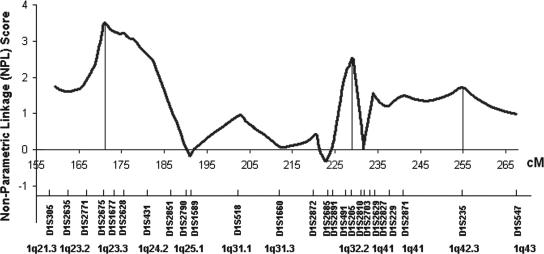
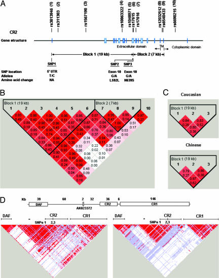
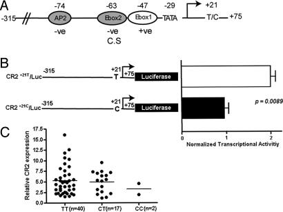
A genomic region on distal mouse chromosome 1 and its syntenic human counterpart 1q23-42 show strong evidence of harboring lupus susceptibility genes. We found evidence of linkage at 1q32.2 in a targeted genome scan of 1q21-43 in 126 lupus multiplex families containing 151 affected sibpairs (nonparametric linkage score 2.52, P = 0.006). A positional candidate gene at 1q32.2, complement receptor 2 (CR2), is also a candidate in the murine Sle1c lupus susceptibility locus. To explore its role in human disease, we analyzed 1,416 individuals from 258 Caucasian and 142 Chinese lupus simplex families and demonstrated that a common three-single-nucleotide polymorphism CR2 haplotype (rs3813946, rs1048971, rs17615) was associated with lupus susceptibility (P = 0.00001) with a 1.54-fold increased risk for the development of disease. Single-nucleotide polymorphism 1 (rs3813946), located in the 5' untranslated region of the CR2 gene, altered transcriptional activity, suggesting a potential mechanism by which CR2 could contribute to the development of lupus. Our findings reveal that CR2 is a likely susceptibility gene for human lupus at 1q32.2, extending previous studies suggesting that CR2 participates in the pathogenesis of systemic lupus erythematosus.
Conflict of interest statement
The authors declare no conflict of interest.
Figures
Similar articles
-
Association of Complement Receptor 2 Gene Polymorphisms with Susceptibility to Osteonecrosis of the Femoral Head in Systemic Lupus Erythematosus.Biomed Res Int. 2016;2016:9208035. doi: 10.1155/2016/9208035. Epub 2016 Jun 30. Biomed Res Int. 2016. PMID: 27446959 Free PMC article.
-
Cr2, a candidate gene in the murine Sle1c lupus susceptibility locus, encodes a dysfunctional protein.Immunity. 2001 Nov;15(5):775-85. doi: 10.1016/s1074-7613(01)00228-x. Immunity. 2001. PMID: 11728339
-
Transcriptional effects of a lupus-associated polymorphism in the 5' untranslated region (UTR) of human complement receptor 2 (CR2/CD21).Mol Immunol. 2012 Oct;52(3-4):165-73. doi: 10.1016/j.molimm.2012.04.013. Epub 2012 Jun 4. Mol Immunol. 2012. PMID: 22673213 Free PMC article.
-
Update on human systemic lupus erythematosus genetics.Curr Opin Rheumatol. 2004 Sep;16(5):513-21. doi: 10.1097/01.bor.0000132648.62680.81. Curr Opin Rheumatol. 2004. PMID: 15314487 Review.
-
Current advances in the human lupus genetics.Curr Rheumatol Rep. 2004 Oct;6(5):391-8. doi: 10.1007/s11926-004-0014-3. Curr Rheumatol Rep. 2004. PMID: 15355752 Review.
Cited by
-
A CD21 low phenotype, with no evidence of autoantibodies to complement proteins, is consistent with a poor prognosis in CLL.Oncotarget. 2015 Oct 20;6(32):32669-80. doi: 10.18632/oncotarget.5404. Oncotarget. 2015. PMID: 26452134 Free PMC article.
-
A loss-of-function variant of PTPN22 is associated with reduced risk of systemic lupus erythematosus.Hum Mol Genet. 2009 Feb 1;18(3):569-79. doi: 10.1093/hmg/ddn363. Epub 2008 Nov 3. Hum Mol Genet. 2009. PMID: 18981062 Free PMC article.
-
Pathways: Strategies for susceptibility genes in SLE.Autoimmun Rev. 2010 May;9(7):473-6. doi: 10.1016/j.autrev.2010.02.003. Epub 2010 Feb 8. Autoimmun Rev. 2010. PMID: 20144911 Free PMC article. Review.
-
Regulatory Architecture of the RCA Gene Cluster Captures an Intragenic TAD Boundary, CTCF-Mediated Chromatin Looping and a Long-Range Intergenic Enhancer.Front Immunol. 2022 Jun 13;13:901747. doi: 10.3389/fimmu.2022.901747. eCollection 2022. Front Immunol. 2022. PMID: 35769482 Free PMC article.
-
Complement Receptors and Their Role in Leukocyte Recruitment and Phagocytosis.Front Cell Dev Biol. 2021 Feb 11;9:624025. doi: 10.3389/fcell.2021.624025. eCollection 2021. Front Cell Dev Biol. 2021. PMID: 33644062 Free PMC article. Review.
References
-
- Tsao BP. Trends Immunol. 2003;24:595–602. - PubMed
-
- Tsao BP, Cantor RM, Grossman JM, Kim SK, Strong N, Lau CS, Chen C-J, Shen N, Ginzler EM, Goldstein R, et al. Arthritis Rheum. 2002;46:2928–2936. - PubMed
-
- Cantor RM, Yuan J, Napier S, Kono N, Grossman JM, Hahn BH, Tsao BP. Arthritis Rheum. 2004;50:3203–3210. - PubMed
Publication types
MeSH terms
Substances
Grants and funding
LinkOut - more resources
Full Text Sources
Medical
Molecular Biology Databases
