. 2007 Mar 6;104(10):3967-72.
doi: 10.1073/pnas.0611618104. Epub 2007 Feb 27.

CD8 tumor-infiltrating lymphocytes are predictive of survival in muscle-invasive urothelial carcinoma

Affiliations

CD8 tumor-infiltrating lymphocytes are predictive of survival in muscle-invasive urothelial carcinoma

Padmanee Sharma et al. Proc Natl Acad Sci U S A. .

Abstract

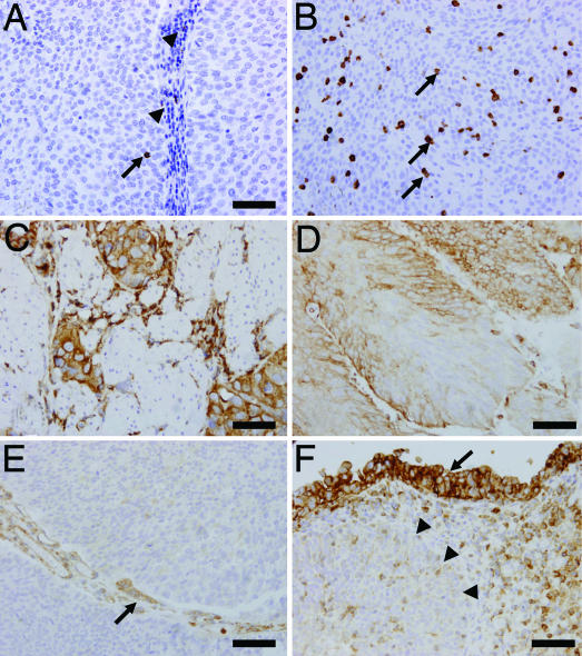
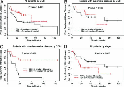
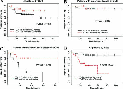
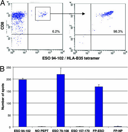
Tumor-infiltrating cytotoxic T lymphocytes (TILs), including CD8 TILs, have been associated with favorable clinical outcomes in multiple tumor types. Tumor-infiltrating CD8 T cells and major histocompatibility complex (MHC) class I expression in urothelial carcinoma (UC) have not been previously reported. Most immune responses are mediated by local cytotoxic lymphocytes (CD8 T cells), which can eradicate tumor cells by recognizing tumor-associated antigens presented by MHC class I molecules. Here we analyzed the presence of intratumoral CD8 T cells, the expression of MHC class I antigen, and the expression of the NY-ESO-1 tumor antigen in UC samples and correlated our findings with clinical outcome. Immunohistochemical staining for intratumoral CD8 T cells in tissue samples from 69 patients with UC showed that patients with advanced UC (pT2, pT3, or pT4) and higher numbers of CD8 TILs within the tumor (> or =8) had better disease-free survival (P < 0.001) and overall survival (P = 0.018) than did patients with similar-staged UC and fewer intratumoral CD8 TILs. We conclude that the extent of intratumoral CD8 TILs is an important prognostic indicator in advanced UC.


Conflict of interest statement

The authors declare no conflict of interest.

Figures

Fig. 1.
Fig. 1.
TILs and expression of MHC class I antigen in representative UC tumor samples. (A) An intratumoral CD8 TIL (arrow) accompanied by stromal CD8 lymphocyte infiltration (arrowheads). (B) Abundant CD8 TILs (arrows) infiltrating a solid tumor with no stromal component. (C) Muscle-invasive case showing heterogeneous expression of class I MHC. (D) Homogeneous expression of class I MHC in another tumor. (E) Class I MHC are not expressed by cancer cells, but are expressed by endothelial cells (arrow). (F) Down-regulation of class I MHC expression associated with cancer invasion. Class I MHC is expressed in the mucosal component (arrow), but not in the invasive component (arrowheads). (Scale bar, 50 μm.)
Fig. 2.
Fig. 2.
Kaplan–Meier plots of DFS according to CD8 TILs and disease stage. P values are from log-rank tests. (A) DFS in all patients according to CD8 TILs <8 (n = 35) vs. CD8 cells ≥8 (n = 34). (B) DFS in patients with early stage (pT1/pTa) disease by CD8 cells <8 (n = 23) vs. CD8 cells ≥8 (n = 15). (C) DFS in patients with advanced (pT2/pT3/pT4) disease by CD8 cells <8 (n = 12) vs. CD8 cells ≥8 (n = 19). (D) DFS in all patients according to disease stage: early stage (T1/Ta; n = 38) vs. advanced (T2/T3/T4; n = 31).
Fig. 3.
Fig. 3.
Kaplan–Meier plots of OS according to CD8 TILs and disease stage. P values are from log-rank tests. (A) OS in all patients according to CD8 TILs <8 (n = 35) vs. CD8 cells ≥8 (n = 34). (B) OS in patients with early stage (T1/Ta) disease by CD8 cells <8 (n = 23) vs. CD8 cells ≥8 (n = 15). (C) OS in patients with advanced (T2/T3/T4) disease by CD8 cells <8 (n = 12) vs. CD8 cells ≥8 (n = 19). (D) OS in all patients according to disease stage: early stage (T1/Ta; n = 38) vs. advanced (T2/T3/T4; n = 31).
Fig. 4.
Fig. 4.
Clonal isolation of CD8 T cells against NY-ESO-1 from the peripheral blood of patient 17. (A) CD8 T cells were stimulated with NY-ESO-1 peptide 79–108 (GARGPESRLLEFYLAMPFATPMEAELARRS), and the resulting polyclonal line was stained with tetramers of synthetic HLA-B*3501/NY-ESO-1 94–102 complexes and with anti-CD8 antibody (Left). Percentages indicate double-positive cells, which were sorted by flow cytometry; several clones were derived and confirmed by tetramer staining (Right). (B) ELISPOT assay showing number of spots corresponding to IFN-γ secretion by cells specific for NY-ESO-1 94–102. These cells also reacted against the 30-mer peptide NY-ESO-1 79–108 and the full-length NY-ESO-1 encoded from recombinant fowlpox virus (FP-ESO), but not against control peptide (FP) or vector (NO PEPT).

Similar articles

Cited by

References

    1. Alexandroff AB, Jackson AM, O'Donnell MA, James K. Lancet. 1999;353:1689–1694. - PubMed
    1. Ponticello A, Perna F, Maione S, Stradolini M, Testa G, Terrazzano G, Ruggerio G, Malerba M, Sanduzzi A. Respir Med. 2004;98:509–514. - PubMed
    1. Sato E, Olson SH, Ahn J, Kepper J, Odunsi T, Ritter G, Lele S, Chen YT, Ohtani H, Old LJ, Odunsi K, et al. Proc Natl Acad Sci USA. 2005;102:18538–18543. - PMC - PubMed
    1. Zhang L, Conejo-Garcia JR, Katsaros D, Gimott PA, Massobrio M, Regnani G, Makrigiannakis A, Gray H, Schlienger K, Liebman MN, et al. N Engl J Med. 2003;348:203–213. - PubMed
    1. Schumacher K, Haensch W, Roefzaad C, Schlag PM. Cancer Res. 2001;61:3932–3936. - PubMed

Publication types

MeSH terms