. 2007 Mar 6;104(10):3984-9.
doi: 10.1073/pnas.0700191104. Epub 2007 Feb 27.

Invariant NKT cells sustain specific B cell responses and memory

Affiliations

Invariant NKT cells sustain specific B cell responses and memory

Grazia Galli et al. Proc Natl Acad Sci U S A. .

Abstract

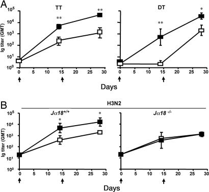
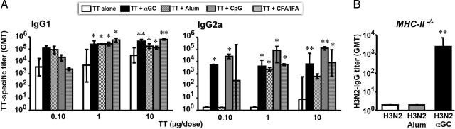
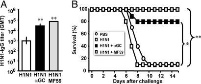
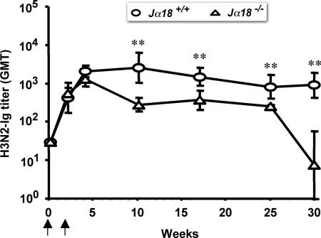
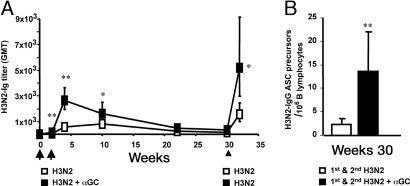
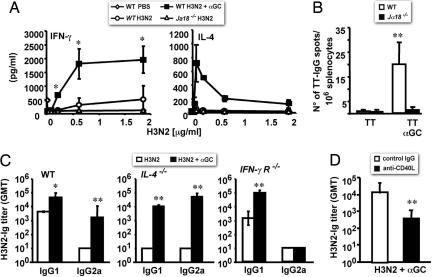
Invariant natural killer T (iNKT) cells are innate-like lymphocytes recognizing CD1d-restricted glycolipid antigens, such as alpha-galactosylceramide (alphaGC). We assessed whether iNKT cells help B lymphocyte responses and found that mice immunized with proteins and alphaGC develop antibody titers 1-2 logs higher than those induced by proteins alone. Activation of iNKT cells enhances protection against infections such as influenza and elicits higher frequencies of memory B cells and higher antibody responses to booster immunizations. Protein vaccination with alphaGC, but not with conventional adjuvants, elicits IgG responses in mice lacking MHC class II molecules, demonstrating that iNKT cells can substitute for CD4(+) T cell help to B cells. Interestingly, the decay of circulating antibodies is faster in mice lacking iNKT cells. These findings point to a homeostatic role for iNKT cells on critical features of the antibody response such as immunity and B cell memory.


Conflict of interest statement

The authors declare no conflict of interest.

Figures

Fig. 1.
Fig. 1.
Activated iNKT cells help antibody responses to bacterial and viral proteins in vivo. Groups of four to six mice were immunized twice (arrows) with the indicated proteins, given alone (□) or with αGC (■). (A) GMTs (and 95% C.I.) of circulating antigen-specific antibodies in C57BL/6 WT mice immunized with TT or DT with or without αGC. (B) GMTs and 95% C.I. of antigen-specific antibodies in sera from C57BL/6 mice bearing (Jα18+/+) or lacking (Jα18−/−) iNKT cells, immunized with H3N2 or H3N2 plus αGC. Results are from one experiment of six (TT), two (DT), or four (H3N2). Asterisks indicate statistically different values between mice immunized with the protein alone and mice immunized with the protein and αGC (∗, P < 0.05; ∗∗, P < 0.01).
Fig. 2.
Fig. 2.
Activated iNKT cells help T-dependent antibody responses like conventional adjuvants and can substitute for MHC-II-restricted T lymphocytes. (A) IgG1 and IgG2a subclass distribution of circulating TT-specific antibodies in C57BL/6 mice 2 weeks after two immunizations with increasing doses of TT alone, TT plus αGC, TT plus Alum, TT plus CpG, TT plus complete Freund's adjuvant (CFA)/incomplete Freund's adjuvant (IFA). (B) GMTs and 95% C.I. of H3N2-specific IgG in sera from C57BL/6 MHC-II−/− mice 2 weeks after two immunizations with H3N2 alone, H3N2 plus Alum, or H3N2 plus αGC. Results are from one experiment representative of three, in which 3–10 mice per group were tested. Asterisks indicate statistically different values between mice immunized with the protein alone and mice immunized with the protein and the given adjuvant (∗, P < 0.05; ∗∗, P < 0.01).
Fig. 3.
Fig. 3.
iNKT cell activation enhances the protective efficacy of a subunit influenza vaccine. (A) GMTs and 95% C.I. of circulating H1N1-specific IgG in C57BL/6 mice 2 weeks after two immunizations with H1N1 alone, H1N1 plus αGC, or H1N1 plus MF59. (B) Survival curves of the mice described in A and control mice injected with PBS alone after intranasal challenge with the influenza virus A/WS/33. Results are from one experiment representative of two in which 10 mice per group were tested. Asterisks indicate statistically different values between mice immunized with H1N1 alone and mice immunized with H1N1 plus αGC or H1N1 plus MF59 (∗, P < 0.05; ∗∗, P < 0.002).
Fig. 4.
Fig. 4.
iNKT cells sustain serum antibody titers. Decay of serum antibodies in C57BL/6 mice bearing (Jα18+/+) or lacking (Jα18−/−) iNKT cells immunized (arrows) with H3N2 alone. Results are from one experiment representative of three in which five mice per group were tested. Asterisks indicate statistically different values (P < 0.03) between the two groups of mice.
Fig. 5.
Fig. 5.
Activated iNKT cells enhance B cell memory. (A) GMTs and 95% C.I. of H3N2-specific antibodies in C57BL/6 WT mice before and after two immunizations (arrows) with H3N2 or H3N2 plus αGC and a subsequent boost (arrowhead) with H3N2 alone. (B) Numbers (average ± SD) of H3N2-IgG ASC precursors per 106 B lymphocytes found on week 30 in the spleens from two groups of mice primed as in A. Results are from one experiment representative of two, in which seven mice per group were analyzed. Asterisks indicate statistically different values (∗, P < 0.05; ∗∗, P < 0.01) between the two groups of mice.
Fig. 6.
Fig. 6.
CD40/CD40L interactions are required for iNKT cell help to antibody responses, whereas IL-4 and IFN-γR are dispensable. (A) IFN-γ and IL-4 released in response to H3N2 in vitro by splenocytes obtained from WT and Jα18−/− C57BL/6 injected with PBS, H3N2, or H3N2 plus αGC. Shown are average values (±SD) obtained by analyzing three mice per group. (B) Numbers (average ± SD) of PCs producing TT-specific IgG in the BM from WT and Jα18−/− C57BL/6 mice (six mice per group) 8 weeks after the second immunization with TT alone or TT plus αGC. (C) IgG1/IgG2a subclass distribution of H3N2-specific serum antibodies in WT C57BL/6 mice or in congenic mice lacking either IL-4 (IL-4−/−) or the IFN-γR (IFN-γR−/−), 2 weeks after two immunizations with H3N2 or H3N2 plus αGC. Shown are GMTs with 95% C.I. obtained analyzing three mice per group. (D) GMTs with 95% C.I. of H3N2-specific antibodies found on day 14 in sera from C57BL/6 WT mice (five mice per group) treated on days −1, 0, and +1 with saturating amounts of a neutralizing anti-CD40L mAb or control IgG and immunized on day 0 with H3N2 plus αGC. Results from one experiment representative of two are shown. Asterisks indicate statistically different values (∗, P < 0.05; ∗∗, P < 0.01) between groups of mice of the same genetic background immunized with the protein alone or the protein plus αGC.

References

    1. Rajewsky K. Nature. 1996;381:751–758. - PubMed
    1. Trinchieri G. Nat Rev Immunol. 2003;3:133–146. - PubMed
    1. Kaisho T, Akira S. Curr Mol Med. 2003;3:373–385. - PubMed
    1. Pasare C, Medzhitov R. Nature. 2005;438:364–368. - PubMed
    1. Ruprecht CR, Lanzavecchia A. Eur J Immunol. 2006;36:810–816. - PubMed

Publication types