. 2007 Mar 6;104(10):3996-4001.
doi: 10.1073/pnas.0609028104. Epub 2007 Feb 23.

Endogenous IL-12 triggers an antiangiogenic program in melanoma cells

Affiliations

Endogenous IL-12 triggers an antiangiogenic program in melanoma cells

Irma Airoldi et al. Proc Natl Acad Sci U S A. .

Abstract

The IL12RB2 gene acts as a tumor suppressor in human B cell malignancies. Indeed, Il12rb2 knockout (KO) mice develop spontaneously B cell tumors, but also lung epithelial tumors. This latter phenotype may be related to (i) impairment of host IL-12-mediated immunosurveillance and/or (ii) IL-12 inability to inhibit directly the growth of IL-12 unresponsive malignant cells. To address this issue, we transplanted IL-12R(+) B16 melanoma cells into syngeneic Il12rb2 KO mice with the following rationale: (i) these mice have severe defects in IFN-gamma production, as well as in cytotoxic T lymphocyte and natural killer cell cytotoxicity, and (ii) they produce but do not use IL-12 that can potentially bind to and target tumor cells only. Il12rb2 KO mice displayed higher endogenous serum levels of IL-12 and developed smaller B16 tumors than WT animals. These tumors showed reduced proliferation, increased apoptosis, and defective microvessel formation related to down-regulated expression of a set of proangiogenic genes previously unrelated to IL-12. Such effects depended on direct activity of endogenous IL-12 on tumor cells in KO mice, and hydrodynamic delivered IL-12 caused further reduced tumorigenicity of B16 cells in these mice. A previously undescribed mechanism of the IL-12 antitumor activity has been here identified and characterized.


Conflict of interest statement

The authors declare no conflict of interest.

Figures

Fig. 1.
Fig. 1.
Characterization and tumorigenicity of B16 cells and IL12p70 serum level in KO mice. (A) Expression of IL12rb1 and b2 chains in B16 melanoma cells. (Left) IL12rb1 surface expression in B16 cells, as assessed by flow cytometry. Open profile: IL12rb1 staining; dark profile: isotype matched mAb staining. (Right) IL12rb2 expression in B16 cells, as assessed by RT-PCR. MW, molecular weight; NC, negative control (water in the place of cDNA); PC, positive control (3T3 cell line); B16 cells, B16 melanoma cells (B16). On the right, the expected mw of the amplified band is shown. (B) Quantitative determination of IL-12p70 concentrations in sera from 10 KO mice, as assessed by ELISA. (C) Volume of s.c. tumors grown in KO and WT animals 17 days (Left) and 7 days (Right) after B16 cell inoculation. The differences in size between tumors removed from KO and WT mice either 17 days or 7 days after B16 cell inoculation were evaluated by Mann–Whitney U test. Boxes indicate values between the 25th and 75th percentiles, whisker lines represent highest and lowest values for each group. Horizontal lines represent median values.
Fig. 2.
Fig. 2.
Histological feature and vascularity of B16 tumors in KO and WT mice. (A) Histological feature of B16 tumors grown after 7 days in WT (Aa and Ac) and KO (Ab and Ad) mice. s.c. injection of B16 cells in both WT (Aa) and KO (Ab) mice gives rise to solid tumor masses showing a monomorphous pattern of small round neoplastic cells. However, differently from tumors developed in WT mice, those from KO mice (Ab) are frequently altered by extensive areas of ischemic-hemorrhagic necrosis (N). This condition is associated with a deficiency in microvessel network (Ad) which, by contrast, seems to be well developed in tumors from WT mice (Ac). (Magnification: ×200 in Aa–Ad.) (B) Vascular network in B16 tumors grown after 7 days in WT and KO mice. The numerous microvessels supporting tumor developed in WT mice are endowed with complete and robust basement membrane, evidenced by laminin immunostaining (Ba), and with pericyte covering, evidenced by α-sma immunostaining (Bc). By contrast, in tumor from KO mice, both laminin deposition (Bb) and pericyte covering (Bd) are dramatically compromised. Furthermore, the production and perivascular accumulation of tenascin-C is prominent in tumor from WT mice (Be) and deficient in tumor from KO mice (Bf).
Fig. 3.
Fig. 3.
In vitro characterization of IL12rb2+ and IL12rb2 B16 clones. (A) Expression of IL12rb2 mRNA in B16 cells, and in IL12rb2+ and IL12rb2 B16 clones, as assessed by RT-PCR. MW, molecular weight; NC, negative control (water in the place of cDNA); PC, positive control (3T3 cell line); four different IL12rb2+ B16 clones (clones 1 to 4) and four different IL12rb2 B16 clones (from clones 5 to 8). On the right, the expected mw of the amplified band is shown. (B) Angiogenic activity of supernatants from one representative IL12rb2+ (clone 1) and one IL12rb2 clone (clone 7) cultured in the presence or absence of mrIL12. CAMs treated with sponges loaded with the conditioned medium from the IL12rb2+ clone were surrounded by allantoic vessels developing radially toward the implant in a “spoked-wheel” pattern (Ba). When medium from IL12rb2+ clone cultured with mrIL12 was tested, a significant reduction of the angiogenic response was evident (Bb). Supernatants from IL12rb2 B16 clone cultured in the presence (Bd) or absence (Bc) of mrIL12 elicited an angiogenic response superimposable to that induced by medium from the untreated IL-12rb2+ B16 clone (see Ba). One representative experiment is shown. (Original magnification: ×50.) (C) Volume of s.c. tumor grown in KO animals 12 days after IL12rb2+ or IL12rb2 B16 clone inoculation. (Left) Differences in size of tumors formed by each individual IL12rb2+ and IL12rb2 B16 clone is shown. Five animals were inoculated with each clone. Whisker lines represent highest and lowest values for each group. Horizontal lines represent median values. (Right) Differences between tumors formed by the four pooled IL12rb2+ and the four pooled IL12rb2 B16 clones in KO animals (Left) were evaluated by Mann–Whitney U test. Boxes indicate values between the 25th and 75th percentiles, whisker lines represent highest and lowest values for each group. Horizontal lines represent median values. (D) Mouse Angiogenesis RT-PCR-Array on tumors explanted from KO animals 12 days after IL12rb2+ and IL12Rb2 B16 clone inoculation. Columns represent fold differences of individual gene expression between tumors formed by the IL12rb2+ clone 1 and IL12rb2 clone 5. Upper columns (>1.00) represent genes whose expression is up-regulated, lower columns (<1.00) genes whose expression is down-regulated. One experiment representative of the two performed with similar results using two different IL12rb2+ clones and two different IL12rb2 clones is shown.
Fig. 4.
Fig. 4.
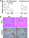
Size and immunohistological features of tumors formed in WT vs. KO mice by an IL12rb2 and an IL12rb2+ B16 clone 7 days after inoculation. (A) The difference in size between IL12rb2+clone 1 tumors from KO and WT mice was statistically significant (Mann–Whitney U test). In contrast, tumors formed in the two groups of mice by the IL12rb2 B16 clone 7 were of similar size. Boxes indicate values between the 25th and 75th percentiles, whisker lines represent highest and lowest values for each group. Horizontal lines represent median values. (B) Morphological features of tumors grown 7 days after IL12rb2 B16 clone 7 injection in WT and KO mice. Both tumors grown in WT (Ba) and in KO (Bb) mice show frequent mitosis (arrows) without signs of ischemia or necrosis. Their histological pattern is similar to that of tumors produced by B16 cell injection (see Fig. 1). A vigorous and mature microvessel network supports tumor growth in both WT (Bc) and KO (Bd) mice, as revealed by laminin immunostaining. (Magnification: ×400 in B.)
Fig. 5.
Fig. 5.
IL12p70 serum concentration and tumorigenicity of IL12rb2+ and IL12rb2 B16 clones in KO mice injected with IL-12p70 containing plasmid. (A) IL12p70 serum concentration was evaluated 6 h, 3 days, and 7 days after inoculation of IL-12p70 plasmid and B16 clones, as assessed by ELISA. (Left) IL-12p70 serum levels from three different animals injected first with IL-12p70 plasmid and 2 h later with IL-12rb2+ clone 1 (▵, ○, □) or IL-12rb2+ clone 2 (•, *, ■), respectively. (Right) IL12p70 serum levels from six different animals injected first with IL12p70 plasmid and 2 h later with the IL12rb2 B16 clone 5 (black symbols) or IL12rb2 B16 clone 7 (white symbols). (B) Tumors grown 7 days after inoculation of the IL12rb2+ clones 1 and 2 in IL12p70 delivered (IL-12) KO mice were significantly smaller than those formed by the same clones in animals injected with control plasmid (GFP) (Mann–Whitney U test). Boxes indicate values between the 25th and 75th percentiles, whisker lines represent highest and lowest values for each group. Horizontal lines represent median values. In contrast, no differences in size were observed between tumors formed by the IL12rRb2 clone 6 in KO mice inoculated with IL-12 or control plasmid.

Similar articles

Cited by

References

    1. Trinchieri G. Nat Rev Immunol. 2003;3:133–146. - PubMed
    1. Trinchieri G, Pflanz S, Kastelein RA. Immunity. 2003;19:641–644. - PubMed
    1. Hunter CA. Nat Rev Immunol. 2005;5:521–531. - PubMed
    1. Wolf SF, Temple PA, Kobayashi M, Young D, Dicig M, Lowe L, Dzialo R, Fitz L, Ferenz C, Hewick RM, et al. J Immunol. 1991;146:3074–3081. - PubMed
    1. Chua AO, Chizzonite R, Desai BB, Truitt TP, Nunes P, Minetti LJ, Warrier RR, Presky DH, Levine JF, Gately MK, et al. J Immunol. 1994;153:128–136. - PubMed

Publication types