. 2007 Mar 6;104(10):4106-11.
doi: 10.1073/pnas.0608798104. Epub 2007 Feb 23.

Antitumor activity of MLN8054, an orally active small-molecule inhibitor of Aurora A kinase

Affiliations

Antitumor activity of MLN8054, an orally active small-molecule inhibitor of Aurora A kinase

Mark G Manfredi et al. Proc Natl Acad Sci U S A. .

Abstract

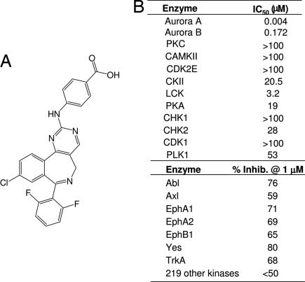
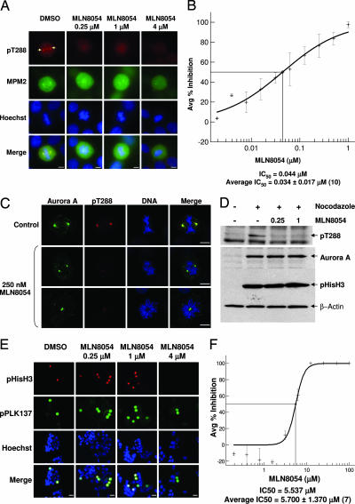
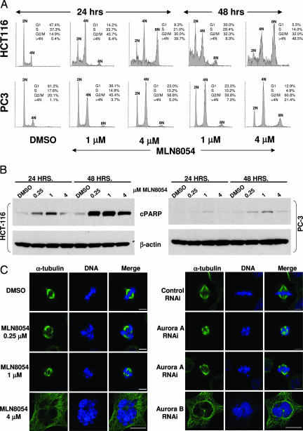
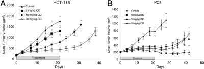
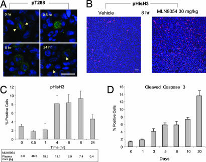
Increased Aurora A expression occurs in a variety of human cancers and induces chromosomal abnormalities during mitosis associated with tumor initiation and progression. MLN8054 is a selective small-molecule Aurora A kinase inhibitor that has entered Phase I clinical trials for advanced solid tumors. MLN8054 inhibits recombinant Aurora A kinase activity in vitro and is selective for Aurora A over the family member Aurora B in cultured cells. MLN8054 treatment results in G(2)/M accumulation and spindle defects and inhibits proliferation in multiple cultured human tumor cells lines. Growth of human tumor xenografts in nude mice was dramatically inhibited after oral administration of MLN8054 at well tolerated doses. Moreover, the tumor growth inhibition was sustained after discontinuing MLN8054 treatment. In human tumor xenografts, MLN8054 induced mitotic accumulation and apoptosis, phenotypes consistent with inhibition of Aurora A. MLN8054 is a selective inhibitor of Aurora A kinase that robustly inhibits growth of human tumor xenografts and represents an attractive modality for therapeutic intervention of human cancers.


Conflict of interest statement

Conflict of interest statement: The authors of this paper, who are employees of Millennium Pharmaceuticals, Inc., are stock holders in the company.

Figures

Fig. 1.
Fig. 1.
MLN8054 is a potent and selective inhibitor of Aurora A kinase. (A) Chemical structure of 4-{[9-chloro-7-(2,6-difluorophenyl)-5H-pyrimido[5,4-d][2]benzazepin-2-yl]amino}-benzoic acid. (B) IC50 values of MLN8054 against recombinant Aurora A, Aurora B, and a panel of other selected kinases. Kinase activity was assessed by using radioactive FlashPlate assays as described in Methods. Percent inhibition for 226 kinases was determined at 1 μM MLN8054 as described in Methods.
Fig. 2.
Fig. 2.
MLN8054 selectively inhibits Aurora A over Aurora B in cultured human tumor cells. (A) Representative immunofluorescent images of HCT-116 cells treated with DMSO or MLN8054 at various concentrations for 24 h. Cells were stained for pT288 (red), MPM2 (green), and DNA (blue). Overlapping localization is shown in the merged images. Arrows indicate Aurora A autophosphorylation on Thr-288, occurring on the centrosomes. (Scale bars, 5 μm.) (B) An Aurora A pT288 autophosphorylation assay was used to measure inhibition of Aurora A by MLN8054 in HeLa cells. The concentration–response curve was generated by calculating the decrease of Aurora A pT288 fluorescent intensity in MLN8054-treated samples relative to the DMSO-treated controls. (C) Immunofluorescent images of HCT-116 cells treated with DMSO or MLN8054 at 250 nM for 24 h. Cells were stained for Aurora A (green), pT288 (red), and DNA (blue). Overlapping localization is shown in the merged images. (Scale bars, 5 μm.) (D) Western blots of nocodazole-arrested cells treated with various concentrations of MLN8054 probed for pT288, Aurora A, pHisH3, and β-actin. (E) Representative immunofluorescent images of HCT-116 cells treated with DMSO or MLN8054 at various concentrations for 24 h. Cells were stained with pHisH3 (Ser-10) mouse monoclonal antibody (pHisH3; red), phospho-PLK (Ser-137, mitotic cells) rabbit antibody (pPLK137; green), and Hoechst (blue). Overlapping localization is shown in the merged images. (Scale bars, 10 μm.) (F) An Aurora B Ser-10 pHisH3 phosphorylation assay was used to measure the inhibition of Aurora B by MLN8054 in HeLa cells. Concentration–response curves were generated by calculating the ratio of Ser-10 pHisH3-positive cells to total mitotic cells in MLN8054-treated samples relative to the DMSO-treated controls. (B and F) For both curves, the points represent averages of three to four replicate samples ± SD (bars). Bisecting lines reflect compound IC50 values. The IC50 number was calculated from the curves shown. Average IC50 numbers represent the average IC50 (μM) ± SD derived from multiple experiments, indicated by the number in parentheses.
Fig. 3.
Fig. 3.
Low and high concentrations of MLN8054 result in cellular phenotypes consistent with Aurora A and Aurora B inhibition, respectively, and induce apoptosis. (A) DNA profiles of HCT-116 and PC3 cells treated with DMSO or MLN8054 at 1 or 4 μM for 24 or 48 h were evaluated by flow cytometry. 2N, 4N, and 8N reflect relative DNA content and represent diploid, tetraploid, and multinucleated cells, respectively. The percentage of cells in G1, S, G2/M and with >4N DNA content is shown. (B) Detection of apoptosis in HCT-116 and PC3 cells with cleaved PARP using Western blot analysis. Cells were treated with DMSO or MLN8054 at 0.25, 1, or 4 μM for 24 or 48 h. (C) Representative immunofluorescent images of HCT-116 cells treated with DMSO or MLN8054 at 0.25, 1, and 4 μM for 24 h or RNAi for control, Aurora A, or Aurora B. Overlapped images were obtained from cells stained with anti-α-tubulin mouse antibody (tubulin; green) and Hoechst (DNA; blue). (Scale bars: DMSO-treated, 0.25 and 1 μM MLN8054-treated, and Aurora A RNAi-treated cells, 5 μm; 4 μM MLN8054-treated and Aurora B RNAi-treated cells, 20 μm.)
Fig. 4.
Fig. 4.
MLN8054 induces TGI in the HCT-116 colorectal and PC3 prostate tumor xenografts. (A) Nude mice bearing HCT-116 tumors were treated orally QD for 21 consecutive days with either vehicle control (▲) or MLN8054 at doses of 3 (■), 10 (●), or 30 (○) mg/kg. Mean tumor volumes (mm3) ± SEM (n = 8–10/group) are shown from the initiation of treatment (≈200 mm3). (B) Nude mice bearing PC3 tumors were treated orally for 21 consecutive days with vehicle control BID (♦) or MLN8054 at 10 mg/kg BID (■), 30 mg/kg BID (▲), or 30 mg/kg QD (×). Mean tumor volumes (mm3) ± SEM (n = 8–10/group) are shown from the beginning of treatment.
Fig. 5.
Fig. 5.
MLN8054 results in inhibition of Aurora A and accumulation of mitotic cells after a single dose and apoptosis after repeat dosing in HCT-116 human tumor xenografts. (A–C) HCT-116 tumor-bearing nude mice were treated orally with a single 30 mg/kg dose of MLN8054, and tumors were removed at various time points for sectioning and immunostaining. DNA (blue) was stained with DAPI to detect all cells in each imaged field. (A) Autophosphorylation of Aurora A on Thr-288 (red, arrows) was determined within mitotic cells (pHisH3-positive; green). (Scale bars, 20 μm.) (B) Mitotic cells (red) were detected by pHisH3 immunostaining and fluorescent microscopy. Shown are representative fields in vehicle-treated control and MLN8054-treated tumors taken 8 h after dosing. (Scale bars, 50 μm.) (C) The percentage of mitotic cells was evaluated at multiple time points throughout a 24-h period. MLN8054 plasma concentrations at each time point were quantified as described in Methods. (D) HCT-116 tumor-bearing mice were treated orally with 30 mg/kg MLN8054 BID for 20 consecutive days. Apoptosis was measured by immunofluorescent staining of cleaved caspase 3. (C and D) The percentage of mitotic and apoptotic cells was calculated (± SD) by using automated imaging microscopy (n = 3 mice per time point, 5 images per slide, and 2 slides per tumor) and by dividing the number of mitotic or apoptotic cells by the total number of cells (DAPI-positive).

References

    1. Bischoff JR, Anderson L, Zhu Y, Mossie K, Ng L, Souza B, Schryver B, Flanagan P, Clairvoyant F, Ginther C, et al. EMBO J. 1998;17:3052–3065. - PMC - PubMed
    1. Nigg EA. Nat Rev Mol Cell Biol. 2001;2:21–32. - PubMed
    1. Crosio C, Fimia GM, Loury R, Kimura M, Okano Y, Zhou H, Sen S, Allis CD, Sassone-Corsi P. Mol Cell Biol. 2002;22:874–885. - PMC - PubMed
    1. Meraldi P, Honda R, Nigg EA. Curr Opin Genet Dev. 2004;14:29–36. - PubMed
    1. Glover DM, Leibowitz MH, McLean DA, Parry H. Cell. 1995;81:95–105. - PubMed

LinkOut - more resources