. 2007 Mar 6;104(10):4112-7.
doi: 10.1073/pnas.0607738104. Epub 2007 Feb 27.

DgrA is a member of a new family of cyclic diguanosine monophosphate receptors and controls flagellar motor function in Caulobacter crescentus

Affiliations

DgrA is a member of a new family of cyclic diguanosine monophosphate receptors and controls flagellar motor function in Caulobacter crescentus

Matthias Christen et al. Proc Natl Acad Sci U S A. .

Erratum in

  • Proc Natl Acad Sci U S A. 2007 May 1;104(18):7729

Abstract

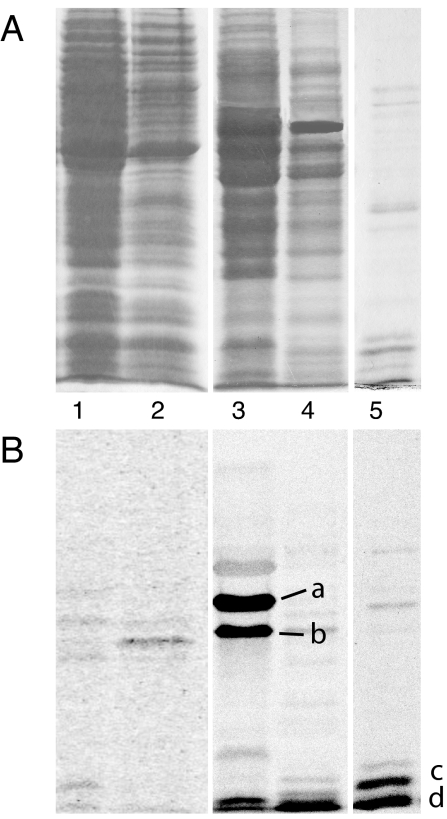
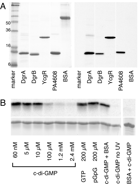
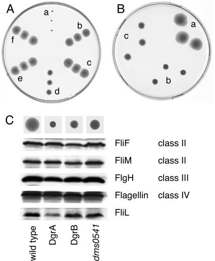
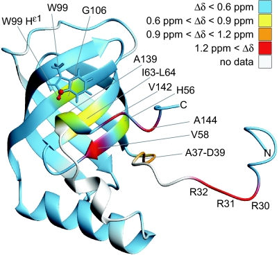
Bacteria are able to switch between two mutually exclusive lifestyles, motile single cells and sedentary multicellular communities that colonize surfaces. These behavioral changes contribute to an increased fitness in structured environments and are controlled by the ubiquitous bacterial second messenger cyclic diguanosine monophosphate (c-di-GMP). In response to changing environments, fluctuating levels of c-di-GMP inversely regulate cell motility and cell surface adhesins. Although the synthesis and breakdown of c-di-GMP has been studied in detail, little is known about the downstream effector mechanisms. Using affinity chromatography, we have isolated several c-di-GMP-binding proteins from Caulobacter crescentus. One of these proteins, DgrA, is a PilZ homolog involved in mediating c-di-GMP-dependent control of C. crescentus cell motility. Biochemical and structural analysis of DgrA and homologs from C. crescentus, Salmonella typhimurium, and Pseudomonas aeruginosa demonstrated that this protein family represents a class of specific diguanylate receptors and suggested a general mechanism for c-di-GMP binding and signal transduction. Increased concentrations of c-di-GMP or DgrA blocked motility in C. crescentus by interfering with motor function rather than flagellar assembly. We present preliminary evidence implicating the flagellar motor protein FliL in DgrA-dependent cell motility control.


Conflict of interest statement

The authors declare no conflict of interest.

Figures

Fig. 1.
Fig. 1.
Isolation of c-di-GMP-binding proteins from C. crescentus. (A) Coomassie blue-stained SDS/polyacrylamide gel with protein fractions used for UV cross-linking with c-[33P]di-GMP. Lane 1, 100.000 × g supernatant; lane 2, 60% ammonium sulfate precipitation; lane 3, 0.4–0.7 M NaCl eluate from Blue Sepharose; lane 4, 0.7–0.9 M NaCl eluate from Blue Sepharose; and lane 5, 125 mM NaCl eluate from GTP-Sepharose column. (B) Autoradiograph of SDS/polyacrylamide gel shown in A. C-di-GMP-binding proteins a, b, and d were identified by MS/MS. Protein c was identified by MS/MS as hypothetical protein CC1599 and was renamed DgrA.
Fig. 2.
Fig. 2.
DgrA is a member of a new family of c-di-GMP-binding proteins. (A) UV cross-linking of purified His6-tagged diguanylate receptor proteins with c-[33P]di-GMP. The following proteins were used: DgrA (CC1599; C. crescentus), DgrB (CC3165; C. crescentus), PA4608 (P. aeruginosa), YcgR (S. typhimurium), and BSA (control). The Coomassie blue-stained gel (Left) and the autoradiograph (Right) are shown. (B) UV cross-linking of 10 μM DgrA in the presence of 60 nM 33P-labeled c-di-GMP. Autoradiograph (Upper) and Coomassie-stained gel (Lower). (Left) Samples were supplemented with increasing concentrations of nonlabeled nucleotides as indicated. (Right) Controls were carried out in the absence of UV irradiation or with BSA.
Fig. 3.
Fig. 3.
DgrA and DgrB are involved in motility control by c-di-GMP. Motility behavior of C. crescentus wild-type strain CB15 and mutants is shown on semisolid agar plates. Three different colonies from independent conjugation experiments are shown. (A) The following strains containing plasmid pUJ142::dgcA or control plasmid pUJ142 were analyzed: CB15/pUJ142::dgcA (a), CB15ΔdgrA/pUJ142::dgcA (b), CB15dgrAW75A/pUJ142::dgcA (c), CB15ΔdgrB/pUJ142::dgcA (d), CB15ΔdgrAΔdgrB/pUJ142::dgcA (e), and CB15/pUJ142 (f). (B) Overexpression of dgrA or dgrB from the lactose promoter (Plac) repressed C. crescentus motility. (a) CB15/pBBR (vector control). (b) CB15/pBBR::dgrA. (c) CB15/pBBR::dgrB. (C) Levels of class II, class III, and class IV structural components of the C. crescentus flagellum were determined by immunoblot analysis for the following strains: CB15/pBBR (wild-type), CB15/pBBR::dgrA (DgrA), CB15/pBBR::dgrB (DgrB), and the extragenic diguanylate receptor motility suppressors CB15dms0541 pBBR::dgrA (dms0541). The motility behavior of each strain is shown on top of the graph.
Fig. 4.
Fig. 4.
Combined amide 1H and 15N shift differences (Δδ) between PA4608 in its free and ligand-bound form. Shift differences are color-coded on the structure of free PA4608 (PDB 1YWU, model 12). Combined chemical shift differences were calculated as Δδ=([(ΔδH)2+(ΔδN/5)2]/2). These data are also shown in SI Fig. 8. Residue Trp99 is shown as sticks, and Nε1 and Hε1 are shown in red to highlight the large Δδ value (1.67 ppm) for these atoms.
Fig. 5.
Fig. 5.
C-di-GMP binding and motility control of DgrA mutants. (A) UV cross-linking of different DgrA mutant proteins with c-[33P]di-GMP. Coomassie blue-stained SDS/PAGE (Upper) and autoradiograph (Lower) with purified wild-type and mutant DgrA proteins (10 μM) are shown. (B) Motility behavior of C. crescentus wild-type CB15 overexpressing different dgrA alleles: CB15/pBBR::dgrA (a), CB15/pBBR::dgrAR11AR12A (b), CB15/pBBR::dgrAD38A (c), CB15/pBBR::dgrAV74A (d), CB15/pBBR::dgrAR11AR12AV74A (e), CB15/pBBR::dgrAW75A (f), and CB15/pBBR (vector control) (g). Three different colonies from independent conjugation experiments are shown.
Fig. 6.
Fig. 6.
Sequence alignment of the c-di-GMP-binding proteins DgrA, DgrB, YcgR, and PA4608 according to the PilZ Pfam entry PF07238. The PilZ domain is highlighted in green. DgrA residues shown to be important for c-di-GMP binding and in vivo function (red) and the positions of intragenic dms suppressor mutations (black) are highlighted above the alignment. Residues of PA4608 with large chemical shift differences upon c-di-GMP binding (blue) are indicated below the alignment.

References

    1. Kolter R, Greenberg EP. Nature. 2006;441:300–302. - PubMed
    1. Jenal U, Malone J. Annu Rev Genet. 2006;40:385–407. - PubMed
    1. Paul R, Weiser S, Amiot NC, Chan C, Schirmer T, Giese B, Jenal U. Genes Dev. 2004;18:715–727. - PMC - PubMed
    1. Ryjenkov DA, Tarutina M, Moskvin OV, Gomelsky M. J Bacteriol. 2005;187:1792–1798. - PMC - PubMed
    1. Schmidt AJ, Ryjenkov DA, Gomelsky M. J Bacteriol. 2005;187:4774–4781. - PMC - PubMed

Publication types

MeSH terms

LinkOut - more resources