. 2007 Mar 6;104(10):4130-5.
doi: 10.1073/pnas.0610167104. Epub 2007 Feb 28.

HIV-1 Vpr function is mediated by interaction with the damage-specific DNA-binding protein DDB1

Affiliations

HIV-1 Vpr function is mediated by interaction with the damage-specific DNA-binding protein DDB1

Bärbel Schröfelbauer et al. Proc Natl Acad Sci U S A. .

Abstract

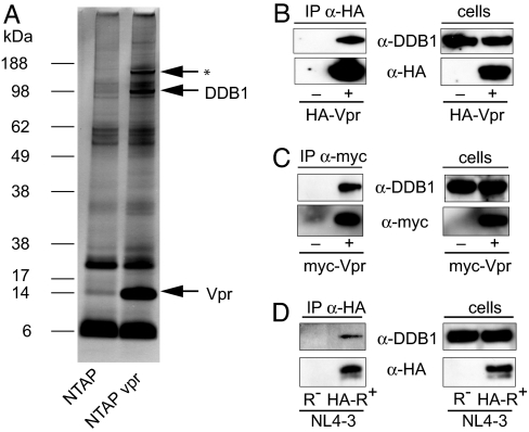
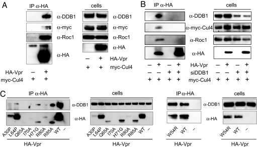
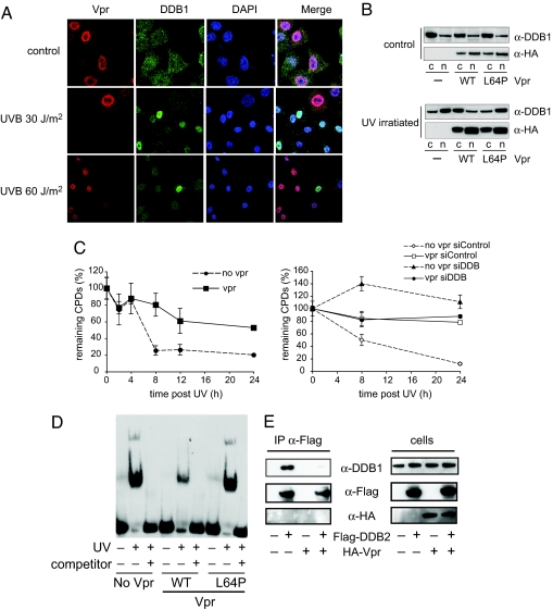
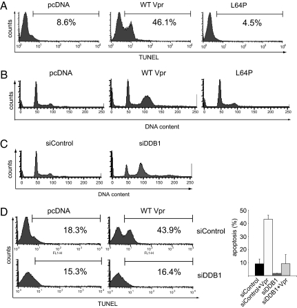
The Vpr accessory protein of HIV-1 induces a response similar to that of DNA damage. In cells expressing Vpr, the DNA damage sensing kinase, ATR, is activated, resulting in G(2) arrest and apoptosis. In addition, Vpr causes rapid degradation of the uracil-DNA glycosylases UNG2 and SMUG1. Although several cellular proteins have been reported to bind to Vpr, the mechanism by which Vpr mediates its biological effects is unknown. Using tandem affinity purification and mass spectrometry, we identified a predominant cellular protein that binds to Vpr as the damage-specific DNA-binding protein 1 (DDB1). In addition to its role in the repair of damaged DNA, DDB1 is a component of an E3 ubiquitin ligase that degrades numerous cellular substrates. Interestingly, DDB1 is targeted by specific regulatory proteins of other viruses, including simian virus 5 and hepatitis B. We show that the interaction with DDB1 mediates Vpr-induced apoptosis and UNG2/SMUG1 degradation and impairs the repair of UV-damaged DNA, which could account for G(2) arrest and apoptosis. The interaction with DDB1 may explain several of the diverse biological functions of Vpr and suggests potential roles for Vpr in HIV-1 replication.


Conflict of interest statement

The authors declare no conflict of interest.

Figures

Fig. 1.
Fig. 1.
Identification of DDB1 as Vpr interacting protein. (A) Protein complexes containing TAP-tagged Vpr from lysates of transfected 293 cells were purified over streptavidin and calmodulin and a portion was visualized on a silver-stained SDS/PAGE. DDB1 and Vpr are indicated with arrows. The three bands were excised and subject to MALDI-TOF mass spectrometry. Two of the bands were identified as DDB1 and Vpr. This identification was reproduced in three independent analyses with separate preparations on different instruments. The third (indicated by the asterisk) was not identified. (B) HA-Vpr was expressed in transfected 293T cells and immunoprecipitated with anti-HA mAb. Coimmunoprecipitated DDB1 was detected on an immunoblot probed with anti-DDB1. (C) Myc-tagged SIVmac Vpr was expressed in transfected cells, immunoprecipitated with anti-Myc mAb and the coimmunoprecipitated DDB1 was detected on an immunoblot. (D) CEMss cells were infected with a replication-competent NL4-3 [NL4-3(HA-Vpr)] engineered to express HA-tagged Vpr or with control Δvpr NL4-3. After 4 days, HA-Vpr was immunoprecipitated with anti-HA mAb. Coimmunoprecipitated DDB1 was detected on an immunoblot probed with anti-DDB1. (B, C, and D Right) Immunoblot analysis of the cell lysates to confirm expression of the relevant proteins.
Fig. 2.
Fig. 2.
Vpr interacts with DDB1-Cul4A E3 ubiquitin ligase complexes. (A Left) HA-Vpr and Myc-Cul4 were expressed in cotransfected 293T cells, and HA-Vpr was immunoprecipitated with anti-HA mAb. Coimmunoprecipitated DDB1, myc-Cul4, and Roc1 were detected on an immunoblot. (Right) Expression of the proteins was confirmed by immunoblot analysis of the cell lysates. (B Left) DDB1 was knocked down by siRNA transfection. To detect the association of Vpr with DDB1 Cul4A and Roc1, Vpr was immunoprecipitated, and coimmunoprecipitated proteins were detected on an immunoblot. (Right) The efficiency of DDB1 knockdown was determined by immunoblot analysis of the cell lysates. (C) HA-Vpr point mutants were expressed in transfected 293T cells. HA-Vpr was immunoprecipitated, and the coimmunoprecipitated DDB1 was detected on an immunoblot (Left). Stability of the mutant Vpr was determined by immunoblot analysis of the cell lysates (second panel from the Left). The W54R Vpr mutant was tested in a separate experiment (Right two panels).
Fig. 3.
Fig. 3.
Vpr recruits the DDB1-Cul4A E3 ubiquitin ligase to target UNG2 for proteasomal degradation. (A) 293T cells were cotransfected with HA-UNG2 expression vector and empty vector, Vpr, or L64P Vpr expression vector. UNG2, Vpr, and tubulin in cell lysates were detected on an immunoblot. (B) DDB1 was knocked down with siRNA. The cells were then transfected with Vpr and UNG2 expression vector. UNG2, Vpr, DDB1, and tubulin were detected on an immunoblot. (C) Cul4 was knocked down in 293T cells. The cells were then transfected with Vpr and UNG2 expression vector. UNG2, Vpr, Cul4A, and tubulin were detected on an immunoblot. (D) 293T cells were cotransfected with UNG2, Vpr, or empty expression vector and myc-DDB1 or W561A myc-DDB1 expression vector. UNG2, Vpr, and DDB1 in the cell lysates were detected on an immunoblot.
Fig. 4.
Fig. 4.
Interaction of Vpr with DDB1 disrupts the UV-DDB complex. (A) Cells were UV irradiated with the indicated dose, and 6 h later Vpr (red) and DDB1 (green) were visualized by immunofluorescence with specific antibody and labeled second antibody. The nuclei (blue) were stained with DAPI. (B) 293T cells were transfected with wild-type or L64P HA-Vpr expression vector or empty vector control. After 2 days, the cells were UV irradiated, and, after an additional 6 h, nuclear and cytoplasmic fractions were prepared. DDB1 and Vpr in the fractions were detected on an immunoblot. (C) HeLa cells transfected with Vpr expression vector or empty vector (Left). The cells were irradiated with 10 J/m2 UV-C and harvested at the indicated time points. The signal from the control cells was subtracted, and counts at t0 were set to 100%. At the indicated time points, genomic DNA was prepared, and the CPD content was measured by ELISA. (Right) The cells were transfected with control or DDB1-specific siRNA before irradiation. The results are representative of at least two repetitions. (D) HeLa cells were transfected with wild-type, L64P Vpr, or empty vector, and, after 2 days, nuclear extracts were prepared. The extracts were incubated with irradiated or unirradiated 5′-biotinylated double stranded oligonucleotide in the presence or absence of a 100-fold molar excess of unlabeled competitor oligonucleotide. Interaction of the oligonucleotide with the UV-DDB complex was detected by electrophoresis mobility shift assay (EMSA). (E) 293T cells were transfected with Flag-DDB2 and HA-Vpr or empty vector. Flag-DDB2 was immunoprecipitated, and coimmunoprecipitated DDB1 and Vpr were detected by immunoblot analysis (Left). An immunoblot on the cell lysates is shown (Right).
Fig. 5.
Fig. 5.
Knockdown of DDB1 relieves Vpr-induced apoptosis. (A) HeLa cells were transfected with wild-type or L64P Vpr expression vector or empty vector. Apoptosis was detected 3 days later by TUNEL. (B) Cells were fixed and stained with propidium iodide and analyzed by FACS. (C) HeLa cells were transfected with control siRNA or siRNA against DDB1. Three days posttransfection, the cells were fixed and stained with propidium iodide. The cell-cycle profiles were analyzed by FACS. (D) HeLa cells were transfected with control siRNA or DDB1-specific siRNA and, 24 h later, with wild-type Vpr. Three days later, apoptosis was measured by TUNEL.

Similar articles

Cited by

References

    1. Lang SM, Weeger M, Stahl-Hennig C, Coulibaly C, Hunsmann G, Muller J, Muller-Hermelink H, Fuchs D, Wachter H, Daniel MM, et al. J Virol. 1993;67:902–912. - PMC - PubMed
    1. Gibbs JS, Lackner AA, Lang SM, Simon MA, Sehgal PK, Daniel MD, Desrosiers RC. J Virol. 1995;69:2378–2383. - PMC - PubMed
    1. Sherman MP, De Noronha CM, Williams SA, Greene WC. DNA Cell Biol. 2002;21:679–688. - PubMed
    1. Muthumani K, Desai BM, Hwang DS, Choo AY, Laddy DJ, Thieu KP, Rao RG, Weiner DB. DNA Cell Biol. 2004;23:239–247. - PubMed
    1. Goh WC, Rogel ME, Kinsey CM, Michael SF, Fultz PN, Nowak MA, Hahn BH, Emerman M. Nat Med. 1998;4:65–71. - PubMed

Publication types