. 2007 Mar 13;104(11):4255-60.
doi: 10.1073/pnas.0700182104. Epub 2007 Mar 6.

Needle-free delivery of macromolecules across the skin by nanoliter-volume pulsed microjets

Affiliations

Needle-free delivery of macromolecules across the skin by nanoliter-volume pulsed microjets

Anubhav Arora et al. Proc Natl Acad Sci U S A. .

Abstract

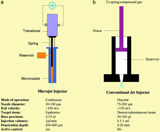
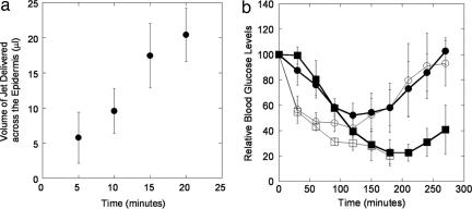
Needle-free liquid jet injectors were invented >50 years ago for the delivery of proteins and vaccines. Despite their long history, needle-free liquid jet injectors are not commonly used as a result of frequent pain and bruising. We hypothesized that pain and bruising originate from the deep penetration of the jets and can potentially be addressed by minimizing the penetration depth of jets into the skin. However, current jet injectors are not designed to maintain shallow dermal penetration depths. Using a new strategy of jet injection, pulsed microjets, we report on delivery of protein drugs into the skin without deep penetration. The high velocity (v >100 m/s) of microjets allows their entry into the skin, whereas the small jet diameters (50-100 mum) and extremely small volumes (2-15 nanoliters) limit the penetration depth ( approximately 200 mum). In vitro experiments confirmed quantitative delivery of molecules into human skin and in vivo experiments with rats confirmed the ability of pulsed microjets to deliver therapeutic doses of insulin across the skin. Pulsed microjet injectors could be used to deliver drugs for local as well as systemic applications without using needles.


Conflict of interest statement

Conflict of interest statement: I.H., J.B., R.R., and R.S. are employees and/or stockholders of StrataGent Life Sciences. S.M. and D.A.F. are scientific advisors and stockholders.

Figures

Fig. 1.
Fig. 1.
Schematic of a pulsed microjet device and conventional jet injector. (a) A pulsed microjet injector comprises a custom-made acrylic micronozzle with final internal diameter in the range of 50–100 μm into which a stainless steel plunger is placed. The plunger is connected to a piezoelectric crystal, which is activated by a custom-designed pulse generator. Activation of the piezoelectric crystal pushes the plunger forward, thereby creating a microjet. Deactivation of the crystal moves the plunger back, and the liquid from the reservoir replenishes displaced liquid. (b) A conventional jet injector comprises a plastic nozzle into which a plunger is placed. The plunger is connected to a compressed spring or compressed gas chamber. Release of compressed spring or gas pushes the plunger to generate a jet. Most commercially available jet injectors are single-use devices (disposable, single-use nozzles attached to a nondisposable device). Typical operating parameters for both types of injectors are listed for the purpose of comparison.
Fig. 2.
Fig. 2.
Performance characteristics of the pulsed microjet injector. (a) Dependence of microjet volume on voltage applied across the piezoelectric crystal. A microjet volume of 15 nl was used for most experiments reported in this study. (b) Dependence of total microjet volume ejected in air as a function of time. The device was operated at a voltage of 140 V across the crystal at a frequency of 1 Hz (n = 3; error bars correspond to SD).
Fig. 3.
Fig. 3.
Penetration of microjets into gel and human skin in vitro. (a) Penetration of microjets into 0.4% wt/vol agarose gel. Microjet was operated at 140 V and 1 Hz. Images represent stills from a video. (bi) Dispersion of dye after delivery by microjet for ≈30 min. (bii) Penetration of conventional jet into 0.4% wt/vol agarose gel delivered by Vitajet 3 (nozzle diameter, 177 μm; velocity >150 m/s) (injection volume of 35 μl). (c) Confocal microscopy pseudocolor images showing penetration of pulsed microjets into full-thickness human skin in vitro (1 μl/min, 1 Hz) (injection volume of 35 μl). (d) Optical images of penetration of conventional jet into human skin in vitro. Jets were delivered from Vitajet 3 (nozzle diameter, 177 μm; velocity >150 m/s). (Upper) Top view. (Lower) Cross-sectional view (injection volume of 35 μl).
Fig. 4.
Fig. 4.
Penetration of microjets into human skin in vitro. The image shows the intact structure of corneocytes around the injection site (bright spot at the center). The image was taken 15–30 min postinjection. (Scale bar, 200 μm.)
Fig. 5.
Fig. 5.
Transdermal delivery mannitol in human skin in vitro and insulin in rat in vivo. (a) Penetration of microjets across human epidermis in vitro (1 μl/min, 1 Hz). Penetration increases linearly with time (n = 3; error bars show SD). (b) Delivery of insulin in Sprague–Dawley rats in vivo (1 μl/min, 1 Hz). Filled squares, microjets delivered for 20 min; filled circles, microjets delivered for 10 min; open circles, s.c. injection of 1.5 units; open squares, conventional jet injection (Vitajet 3, 2 units) (n = 3–5; error bars correspond to SD).

Similar articles

Cited by

References

    1. Kermode M. Health Promot Int. 2004;19:95–103. - PubMed
    1. Varmus H, Klausner R, Zerhouni E, Acharya T, Daar AS, Singer PA. Science. 2003;302:398–399. - PMC - PubMed
    1. Voelker R. J Am Med Assoc. 1999;281:1879–1881. - PubMed
    1. Mitragotri S. Nat Rev Drug Discov. 2006;5:543–548. - PubMed
    1. Levy D, Kost J, Meshulam Y, Langer R. J Clin Invest. 1989;83:2074–2078. - PMC - PubMed

Publication types