

Spiral structure of Escherichia coli HUalphabeta provides foundation for DNA supercoiling
- PMID: 17360520
- PMCID: PMC1838598
- DOI: 10.1073/pnas.0611686104
Spiral structure of Escherichia coli HUalphabeta provides foundation for DNA supercoiling
Abstract
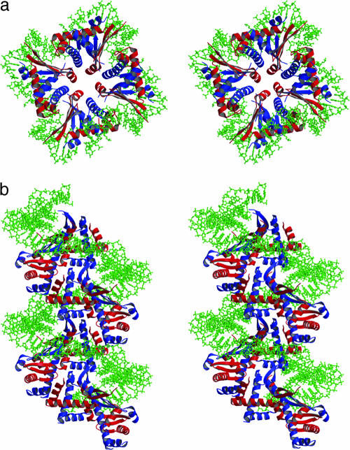
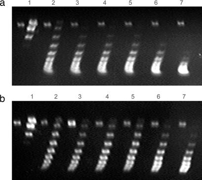
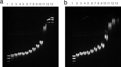
We determined the crystal structure of the Escherichia coli nucleoid-associated HUalphabeta protein by x-ray diffraction and observed that the heterodimers form multimers with octameric units in three potential arrangements, which may serve specialized roles in different DNA transaction reactions. It is of special importance that one of the structures forms spiral filaments with left-handed rotations. A negatively superhelical DNA can be modeled to wrap around this left-handed HUalphabeta multimer. Whereas the wild-type HU generated negative DNA supercoiling in vitro, an engineered heterodimer with an altered amino acid residue critical for the formation of the left-handed spiral protein in the crystal was defective in the process, thus providing the structural explanation for the classical property of HU to restrain negative supercoils in DNA.
Conflict of interest statement
The authors declare no conflict of interest.
Figures


Similar articles
-
DNA reshaping by MukB. Right-handed knotting, left-handed supercoiling.J Biol Chem. 2006 Feb 24;281(8):4606-15. doi: 10.1074/jbc.M504754200. Epub 2005 Dec 20. J Biol Chem. 2006. PMID: 16368697 Free PMC article.
-
Right-handed DNA supercoiling by an octameric form of histone-like protein HU: modulation of cellular transcription.J Biol Chem. 2006 Dec 29;281(52):40144-53. doi: 10.1074/jbc.M605576200. Epub 2006 Oct 23. J Biol Chem. 2006. PMID: 17062578
-
Interaction of the Escherichia coli HU Protein with Various Topological Forms of DNA.Biomolecules. 2021 Nov 19;11(11):1724. doi: 10.3390/biom11111724. Biomolecules. 2021. PMID: 34827722 Free PMC article.
-
DNA condensation in bacteria: Interplay between macromolecular crowding and nucleoid proteins.Biochimie. 2010 Dec;92(12):1715-21. doi: 10.1016/j.biochi.2010.06.024. Epub 2010 Jul 6. Biochimie. 2010. PMID: 20615449 Review.
-
Control of bacterial DNA supercoiling.Mol Microbiol. 1992 Feb;6(4):425-33. doi: 10.1111/j.1365-2958.1992.tb01486.x. Mol Microbiol. 1992. PMID: 1313943 Review.
Cited by
-
Identification of common highly expressed genes of Salmonella Enteritidis by in silico prediction of gene expression and in vitro transcriptomic analysis.Poult Sci. 2019 Jul 1;98(7):2948-2963. doi: 10.3382/ps/pez119. Poult Sci. 2019. PMID: 30953073 Free PMC article.
-
The torsional state of DNA within the chromosome.Chromosoma. 2011 Aug;120(4):323-34. doi: 10.1007/s00412-011-0324-y. Epub 2011 May 13. Chromosoma. 2011. PMID: 21567156 Review.
-
Bacterial chromosome organization and segregation.Annu Rev Cell Dev Biol. 2015;31:171-99. doi: 10.1146/annurev-cellbio-100814-125211. Annu Rev Cell Dev Biol. 2015. PMID: 26566111 Free PMC article. Review.
-
Nonspecific DNA binding and bending by HUαβ: interfaces of the three binding modes characterized by salt-dependent thermodynamics.J Mol Biol. 2011 Jul 8;410(2):241-67. doi: 10.1016/j.jmb.2011.04.001. Epub 2011 Apr 12. J Mol Biol. 2011. PMID: 21513716 Free PMC article.
-
Protein/DNA interactions in complex DNA topologies: expect the unexpected.Biophys Rev. 2016;8(3):233-243. doi: 10.1007/s12551-016-0208-8. Epub 2016 Aug 8. Biophys Rev. 2016. PMID: 27738452 Free PMC article. Review.
References
-
- Johnson RC, Johnson LM, Schmidt J, Gardner JF. In: The Bacterial Chromosome. Higgins NP, editor. Washington, DC: Am Soc Microbiol; 2005. pp. 65–132.
-
- Azam TA, Hiraga S, Ishihama A. Genes Cells. 2000;5:613–626. - PubMed
-
- Claret L, Rouviere-Yaniv J. J Mol Biol. 1997;273:93–104. - PubMed
-
- Castaing B, Zelwer C, Laval J, Boiteux S. J Biol Chem. 1995;270:10291–10296. - PubMed
Publication types
MeSH terms
Substances
Associated data
- Actions
Grants and funding
LinkOut - more resources
Full Text Sources
Other Literature Sources
Molecular Biology Databases
