. 2007 Mar 13;104(11):4383-8.
doi: 10.1073/pnas.0701140104. Epub 2007 Mar 6.

Chemical genetics reveals the requirement for Polo-like kinase 1 activity in positioning RhoA and triggering cytokinesis in human cells

Affiliations

Chemical genetics reveals the requirement for Polo-like kinase 1 activity in positioning RhoA and triggering cytokinesis in human cells

Mark E Burkard et al. Proc Natl Acad Sci U S A. .

Abstract

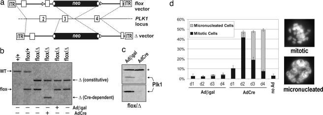
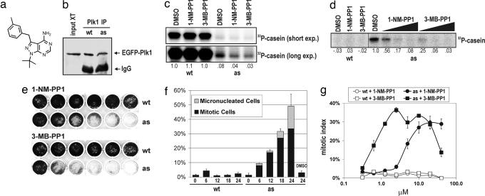
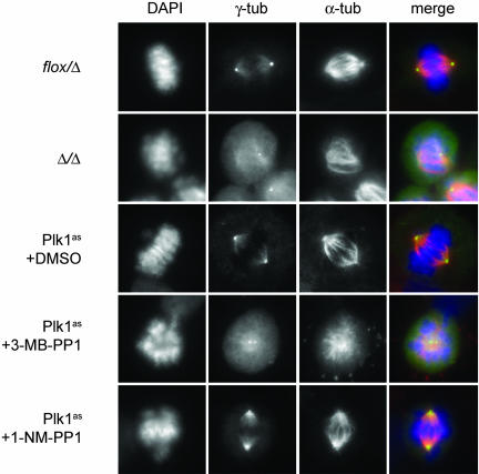
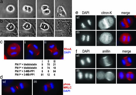
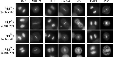
Polo-like kinases (Plks) play crucial roles in mitosis and cell division. Whereas lower eukaryotes typically contain a single Plk, mammalian cells express several closely related but functionally distinct Plks. We describe here a chemical genetic system in which a single Plk family member, Plk1, can be inactivated with high selectivity and temporal resolution by using an allele-specific, small-molecule inhibitor, as well as the application of this system to dissect Plk1's role in cytokinesis. To do this, we disrupted both copies of the PLK1 locus in human cells through homologous recombination and then reconstituted Plk1 activity by using either the wild-type kinase (Plk1(wt)) or a mutant version whose catalytic pocket has been enlarged to accommodate bulky purine analogs (Plk1(as)). When cultured in the presence of these analogs, Plk1(as) cells accumulate in prometaphase with defects that parallel those found in PLK1(Delta/Delta) cells. In addition, acute treatment of Plk1(as) cells during anaphase prevents recruitment of both Plk1 itself and the Rho guanine nucleotide exchange factor (RhoGEF) Ect2 to the central spindle, abolishes RhoA GTPase localization to the equatorial cortex, and suppresses cleavage furrow formation and cell division. Our studies define and illuminate a late mitotic function of Plk1 that, although difficult or impossible to detect in Plk1-depleted cells, is readily revealed with chemical genetics.


Conflict of interest statement

The authors declare no conflict of interest.

Figures

Fig. 1.
Fig. 1.
Conditional inactivation of PLK1 in human cells through gene targeting and Cre-mediated recombination. (a) Diagram of the PLK1 locus and adeno-associated virus vectors used to create floxed (PLK1flox) and null (PLK1Δ) alleles. loxP sites (triangles) and FRT sites (circles) are shown. (b) Southern blot analysis confirming gene replacements and Cre-induced recombination in PLK1flox cells. (c) Extracts from PLK1flox cells infected with Adβgal or AdCre were blotted with antibodies against the C terminus (Upper) or N terminus (Lower) of Plk1. The asterisk denotes a cross-reacting band used to confirm equal loading. (d) Cells infected as in c were scored for the fraction of interphase, mitotic, and micronucleated cells by Hoechst staining and fluorescence microscopy. Error bars denote SEM.
Fig. 2.
Fig. 2.
Reconstitution of Plk1 function with Plk1as. (a) Chemical structure of 3-MB-PP1. (b–d) Nucleotide specificity and analog sensitivity of Plk1wt and Plk1as. Plk1 was immunoprecipitated from mitotic extracts and either immunoblotted (b) or incubated with casein and [γ-32P]ATP (c) or [γ-32P]N6-(benzyl)-ATP (d). Where indicated, 1-NM-PP1 or 3-MB-PP1 was added to the reactions (10 μM, c and left portion of d; 100 nM, 1 μM, and 10 μM, right portion of d). Casein labeling is reported relative to DMSO-treated Plk1wt (c) or Plk1as (d) controls. (e) Plk1as cells are analog-sensitive. Cells were cultured in the presence of 1-NM-PP1 or 3-MB-PP1 (left to right: 0 μM, 0.078 μM, 0.313 μM, 1.25 μM, 5 μM, and 20 μM) for 8 days. (f) Inhibitor-treated Plk1as cells arrest in mitosis. Plk1wt and Plk1as cells were challenged with 10 μM 1-NM-PP1 and sampled at the indicated times. Percentages of mitotic and micronucleated cells were determined as in Fig. 1. (g) 3-MB-PP1 induces mitotic arrest at a 10-fold lower concentration than 1-NM-PP1. Plk1wt (open symbols) and Plk1as cells (closed symbols) were cultured for 18 h in the presence of 1-NM-PP1 (circles) or 3-MB-PP1 (squares). Mitotic indices were determined as above.
Fig. 3.
Fig. 3.
Comparison of early mitotic defects in PLK1-null and Plk1as cells. PLK1flox cells were infected with Adβgal or AdCre for 48 h. Plk1as cells were incubated for 10 h with 10 μM 3-MB-PP1, 10 μM 1-NM-PP1, or DMSO. Cells were stained with antibodies to γ-tubulin (γ-tub) (green) and α-tubulin (α-tub) (red) and analyzed by immunofluorescence microscopy.
Fig. 4.
Fig. 4.
Acute inhibition of Plk1 during anaphase disrupts RhoA localization and blocks the onset of cytokinesis. (a and b) Metaphase Plk1wt (a) and Plk1as (b) cells were treated with 10 μM 3-MB-PP1 (time 0) and followed during progression into anaphase. Although chromatid separation occurred normally, Plk1as cells failed to divide and became binucleated (SI Movies 1 and 2). (c) Plk1 activity is required to localize RhoA at the equatorial cortex. Plk1wt and Plk1as cells were synchronized with monastrol, released for 30 min, and then treated for 20 min with either blebbistatin or 3-MB-PP1. RhoA accumulation at the equatorial cortex was visualized after trichloroacetic acid fixation (21, 27). Anaphase cells (n = 100 per sample) were classified with respect to equatorial RhoA staining and cleavage furrow formation. (d–f) Plk1wt and Plk1as cells were treated with 3-MB-PP1 and stained with antibodies against phosphorylated myosin regulatory light chain (MRLC) (d), citron kinase (citron-K) (e), and anillin (f).
Fig. 5.
Fig. 5.
Plk1 activity recruits both the RhoGEF Ect2 and Plk1 itself to the central spindle. Plk1wt and Plk1as cells were treated as in Fig. 4c. Whereas MKLP1 and CYK-4 localized correctly under all conditions, Ect2 and Plk1 were both absent from the central spindle after Plk1 inhibition.

Similar articles

Cited by

References

    1. van de Weerdt BC, Medema RH. Cell Cycle. 2006;5:853–864. - PubMed
    1. Barr FA, Sillje HH, Nigg EA. Nat Rev Mol Cell Biol. 2004;5:429–441. - PubMed
    1. Peters U, Cherian J, Kim JH, Kwok BH, Kapoor TM. Nat Chem Biol. 2006;2:618–626. - PubMed
    1. McInnes C, Mazumdar A, Mezna M, Meades C, Midgley C, Scaerou F, Carpenter L, Mackenzie M, Taylor P, Walkinshaw M, et al. Nat Chem Biol. 2006;2:608–617. - PubMed
    1. Liu Y, Shreder KR, Gai W, Corral S, Ferris DK, Rosenblum JS. Chem Biol. 2005;12:99–107. - PubMed

Publication types