Triptolide is a traditional Chinese medicine-derived inhibitor of polycystic kidney disease
- PMID: 17360534
- PMCID: PMC1838612
- DOI: 10.1073/pnas.0700499104
Triptolide is a traditional Chinese medicine-derived inhibitor of polycystic kidney disease
Abstract
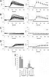
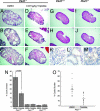
During kidney organogenesis, tubular epithelial cells proliferate until a functional tubule is formed as sensed by cilia bending in response to fluid flow. This flow-induced ciliary mechanosensation opens the calcium (Ca(2+)) channel polycystin-2 (PC2), resulting in a calcium flux-mediated cell cycle arrest. Loss or mutation of either PC2 or its regulatory protein polycystin-1 (PC1) results in autosomal dominant polycystic kidney disease (ADPKD), characterized by cyst formation and growth and often leading to renal failure and death. Here we show that triptolide, the active diterpene in the traditional Chinese medicine Lei Gong Teng, induces Ca(2+) release by a PC2-dependent mechanism. Furthermore, in a murine model of ADPKD, triptolide arrests cellular proliferation and attenuates overall cyst formation by restoring Ca(2+) signaling in these cells. We anticipate that small molecule induction of PC2-dependent calcium release is likely to be a valid therapeutic strategy for ADPKD.
Conflict of interest statement
Conflict of interest statement: C.M.C. and S.S. are currently exploring the commercial implications of these findings and declare a potential conflict of interest.
Figures


Similar articles
-
Human ADPKD primary cyst epithelial cells with a novel, single codon deletion in the PKD1 gene exhibit defective ciliary polycystin localization and loss of flow-induced Ca2+ signaling.Am J Physiol Renal Physiol. 2007 Mar;292(3):F930-45. doi: 10.1152/ajprenal.00285.2006. Epub 2006 Nov 7. Am J Physiol Renal Physiol. 2007. PMID: 17090781 Free PMC article.
-
Triptolide reduces cyst formation in a neonatal to adult transition Pkd1 model of ADPKD.Nephrol Dial Transplant. 2010 Jul;25(7):2187-94. doi: 10.1093/ndt/gfp777. Epub 2010 Feb 4. Nephrol Dial Transplant. 2010. PMID: 20139063 Free PMC article.
-
Adenylyl cyclase 5 links changes in calcium homeostasis to cAMP-dependent cyst growth in polycystic liver disease.J Hepatol. 2017 Mar;66(3):571-580. doi: 10.1016/j.jhep.2016.10.032. Epub 2016 Nov 5. J Hepatol. 2017. PMID: 27826057 Free PMC article.
-
Polycystin 2: A calcium channel, channel partner, and regulator of calcium homeostasis in ADPKD.Cell Signal. 2020 Feb;66:109490. doi: 10.1016/j.cellsig.2019.109490. Epub 2019 Dec 2. Cell Signal. 2020. PMID: 31805375 Free PMC article. Review.
-
Cilia and polycystic kidney disease.Semin Cell Dev Biol. 2021 Feb;110:139-148. doi: 10.1016/j.semcdb.2020.05.003. Epub 2020 May 28. Semin Cell Dev Biol. 2021. PMID: 32475690 Review.
Cited by
-
PTEN-induced partial epithelial-mesenchymal transition drives diabetic kidney disease.J Clin Invest. 2019 Mar 1;129(3):1129-1151. doi: 10.1172/JCI121987. Epub 2019 Feb 11. J Clin Invest. 2019. PMID: 30741721 Free PMC article.
-
Pyrrolidine dithiocarbamate reduces the progression of total kidney volume and cyst enlargement in experimental polycystic kidney disease.Physiol Rep. 2014 Dec 11;2(12):e12196. doi: 10.14814/phy2.12196. Print 2014 Dec 1. Physiol Rep. 2014. PMID: 25501440 Free PMC article.
-
MYC Inhibition Depletes Cancer Stem-like Cells in Triple-Negative Breast Cancer.Cancer Res. 2017 Dec 1;77(23):6641-6650. doi: 10.1158/0008-5472.CAN-16-3452. Epub 2017 Sep 26. Cancer Res. 2017. PMID: 28951456 Free PMC article.
-
Triptolide Inhibits the AR Signaling Pathway to Suppress the Proliferation of Enzalutamide Resistant Prostate Cancer Cells.Theranostics. 2017 Apr 20;7(7):1914-1927. doi: 10.7150/thno.17852. eCollection 2017. Theranostics. 2017. PMID: 28638477 Free PMC article.
-
Novel targets for the treatment of autosomal dominant polycystic kidney disease.Expert Opin Investig Drugs. 2010 Mar;19(3):315-28. doi: 10.1517/13543781003588491. Expert Opin Investig Drugs. 2010. PMID: 20141351 Free PMC article. Review.
References
-
- Gabow PA, Grantham JJ. Polycystic Kidney Disease. Boston: Little, Brown; 1997.
-
- Koulen P, Cai Y, Geng L, Maeda Y, Nishimura S, Witzgall R, Ehrlich BE, Somlo S. Nat Cell Biol. 2002;4:191–197. - PubMed
-
- Anyatonwu GI, Ehrlich BE. J Biol Chem. 2005;280:29488–29493. - PubMed
-
- Igarashi P, Somlo S. J Am Soc Nephrol. 2002;13:2384–2398. - PubMed
-
- Nauli SM, Alenghat FJ, Luo Y, Williams E, Vassilev P, Li X, Elia AE, Lu W, Brown EM, Quinn SJ, et al. Nat Genet. 2003;33:129–137. - PubMed
Publication types
MeSH terms
Substances
Grants and funding
LinkOut - more resources
Full Text Sources
Other Literature Sources
Research Materials
Miscellaneous

