. 2007 Mar 13;104(11):4443-8.
doi: 10.1073/pnas.0610647104. Epub 2007 Mar 7.

RBP-J (Rbpsuh) is essential to maintain muscle progenitor cells and to generate satellite cells

Affiliations

RBP-J (Rbpsuh) is essential to maintain muscle progenitor cells and to generate satellite cells

Elena Vasyutina et al. Proc Natl Acad Sci U S A. .

Abstract

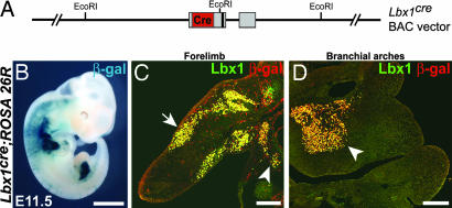
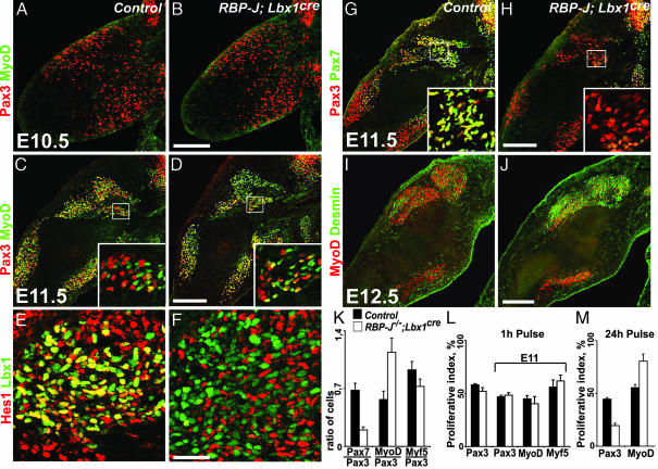
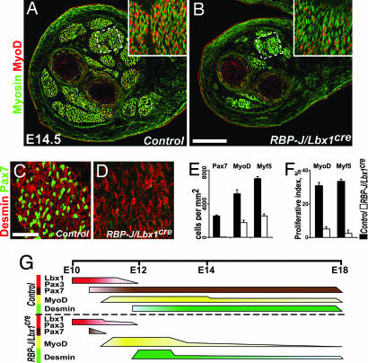
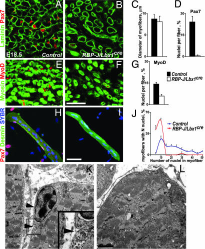
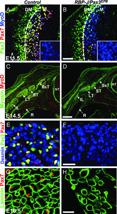
In the developing muscle, a pool of myogenic progenitor cells is formed and maintained. These resident progenitors provide a source of cells for muscle growth in development and generate satellite cells in the perinatal period. By the use of conditional mutagenesis in mice, we demonstrate here that the major mediator of Notch signaling, the transcription factor RBP-J, is essential to maintain this pool of progenitor cells in an undifferentiated state. In the absence of RBP-J, these cells undergo uncontrolled myogenic differentiation, leading to a depletion of the progenitor pool. This results in a lack of muscle growth in development and severe muscle hypotrophy. In addition, satellite cells are not formed late in fetal development in conditional RBP-J mutant mice. We conclude that RBP-J is required in the developing muscle to set aside proliferating progenitors and satellite cells.


Conflict of interest statement

The authors declare no conflict of interest.

Figures

Fig. 1.
Fig. 1.
Recombination introduced by the Lbx1cre transgene. (A) Schematic display of the Lbx1cre transgene. In the modified 144-kb BAC clone, cre-recombinase (red), was fused to the ATG initiation codon of Lbx1 and replaced Lbx1 coding sequences (gray boxes); the vector was used to generate the transgenic Lbx1cre mouse strain. (B–D) Lbx1cre-induced recombination was monitored in embryos (E11.5) carrying the ROSA26R reporter; recombination was assessed by X-Gal staining (B) or by the use of antibodies to detect Lbx1 (green) or β-galactosidase (red) (C and D). (C) Longitudinal section on the forelimb; muscle progenitors in the limb and in the stream moving to the diaphragm are indicated by arrow and arrowhead, respectively. (D) Section of the branchial arches; the arrowhead points to muscle progenitors that subsequently generate the tongue muscle. (Scale bars: B, 2 mm; C and D, 250 μm.)
Fig. 2.
Fig. 2.
Development of myogenic cells in the limb of RBP-J/Lbx1cre mice. Immunohistological analysis of myogenic cells in developing limbs of control and RBP-J/Lbx1cre mice. Myogenic cells were analyzed at E10.5 (A and B), E11.5 (C–H), and E12.5 (I and J) by using the indicated antibodies. Insets (C and D, G and H) show the boxed areas at higher magnification. (K) Ratios of Pax7/Pax3, MyoD/Pax3, and Myf5/Pax3 cells observed at E11.5. (L) Proliferation was assessed by BrdU-labeling; shown are the proportion of Pax3+, MyoD+, and Myf5+ cells labeled 1 h after BrdU injection at E10.5 or E11.5. (M) BrdU was injected at E10.5, and the proportions of Pax3+ or MyoD+ cells that incorporated BrdU were assessed after a 24-h chase. (Scale bars: A–D and G–J, 200 μm; E and F, 50 μm.)
Fig. 3.
Fig. 3.
Differentiated muscle groups in the distal limb of RBP-J/Lbx1cre mice. (A–D) Immunohistological analysis of muscle groups in the distal limb of control (A and C) and RBP-J/Lbx1cre (B and D) mice at E14.5 by using the indicated antibodies. (E) Quantification of Pax7+ and MyoD+ or Myf5+ cells in control and RBP-J/Lbx1cre mice. Shown are the numbers of cells/mm2. (F) Quantification of proliferating MyoD+ and Myf5+ cells in distal limb muscles of control and RBP-J/Lbx1cre mice. BrdU was injected 1 h before the analysis at E14.5. Displayed are the proportions of MyoD+ and Myf5+ cells that incorporated BrdU. (G) Summary of the expression of various markers used to identify myogenic progenitors and differentiating myogenic cells in limbs of control (Upper) and RBP-J/Lbx1cre (Lower) mice during development. Bar thickness indicates cell numbers at particular stages that express the indicated proteins. (Scale bars: A and B, 250 μm; C and D, 50 μm.)
Fig. 4.
Fig. 4.
Satellite cells are absent in the limb of RBP-J/Lbx1cre mice. (A and B) Immunohistological analysis of muscle in distal limbs of control (A) and RBP-J/Lbx1cre (B) mice at E18.5 by using antibodies against laminin (green) and Pax7 (red). (C) Quantification of the myofiber diameter in control and RBP-J/Lbx1cre mice; the outline of myofibers was visualized by using anti-laminin antibodies. (D) Quantification of the number of Pax7+ cells/myofiber in control and RBP-J/Lbx1cre mice. (E and F) Immunohistological analysis of limb muscle in control (E) and RBP-J/Lbx1cre (F) mice at E18.5 by using skeletal muscle-specific myosin (green) and MyoD (red) antibodies. (G) Quantification of the number of MyoD nuclei/myofiber in control and RBP-J/Lbx1cre mice. Immunohistological analyses of single muscle fibers from control (H) and RBP-J/Lbx1cre (I) mice at E18.5 by using desmin (green) and Pax7 (red) antibodies. A nuclear counterstain (SYBR) is shown in blue. (J) Quantification of the number of nuclei/myofiber in control and RBP-J/Lbx1cre mice. (K and L) Ultrastructure of limb muscle from control (K) and RBP-J/Lbx1cre (L) mice at E18.5. In control mice, satellite cells are separated from myofibers by plasma membranes and locate below the basal membrane (arrowheads). In RBP-J mutants, satellite cells were not detected. (Scale bars: A–I, 50 μm; K and L, 2 μm.)
Fig. 5.
Fig. 5.
Myotome and myotome-derived muscle in RBP-J/Pax3cre mice. (A and B) Immunohistological analysis of the dermomyotome and myotome in control and RBP-J/Pax3cre mice at E11.5 by using Pax3 (green), Pax7 (red), and MyoD (blue) antibodies. The stippled lines indicate the border between myotome (M) and dermomyotome (DM). Insets (A and B) display magnifications of the myotome, and demonstrate a higher density of MyoD+ cells in the myotome of mutant mice. (C–F) Analysis of back muscle in control and RBP-J/Pax3cre mice at E14.5 by using the indicated antibodies. (G and H) Analysis of back muscle in control and RBP-J/Pax3cre mice at E18.5 by using laminin (green) and Pax7 (red) antibodies. Neural tube (NT), rib (R), and deep muscles of the back, semispinalis thoracis (SsT), spinalis thoracis (ST), longissimus thoracis (LT), ilicostalis lumborum (IL), are indicated. (Scale bars: A and B, 100 μm; C and D, 250 μm; and E–H, 25 μm.)

Similar articles

Cited by

References

    1. Buckingham M. Curr Opin Genet Dev. 2006;16:525–532. - PubMed
    1. Arnold HH, Braun T. Curr Top Dev Biol. 2000;48:129–164. - PubMed
    1. Buckingham M. Curr Opin Genet Dev. 2001;11:440–448. - PubMed
    1. Parker MH, Seale P, Rudnicki MA. Nat Rev Genet. 2003;4:497–507. - PubMed
    1. Ordahl CP, Le Douarin NM. Development (Cambridge, UK) 1992;114:339–353. - PubMed

Publication types

Substances