. 2007 Mar 13;104(11):4565-70.
doi: 10.1073/pnas.0610335104. Epub 2007 Mar 7.

Impaired NFAT nuclear translocation results in split exhaustion of virus-specific CD8+ T cell functions during chronic viral infection

Affiliations

Impaired NFAT nuclear translocation results in split exhaustion of virus-specific CD8+ T cell functions during chronic viral infection

Paola Agnellini et al. Proc Natl Acad Sci U S A. .

Abstract

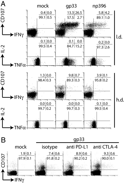
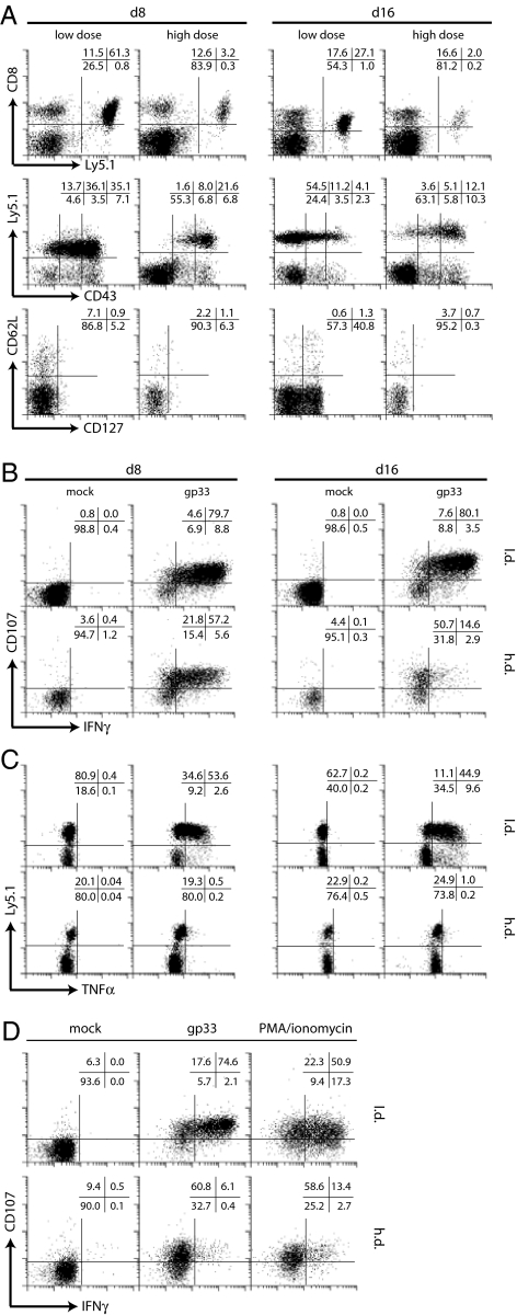
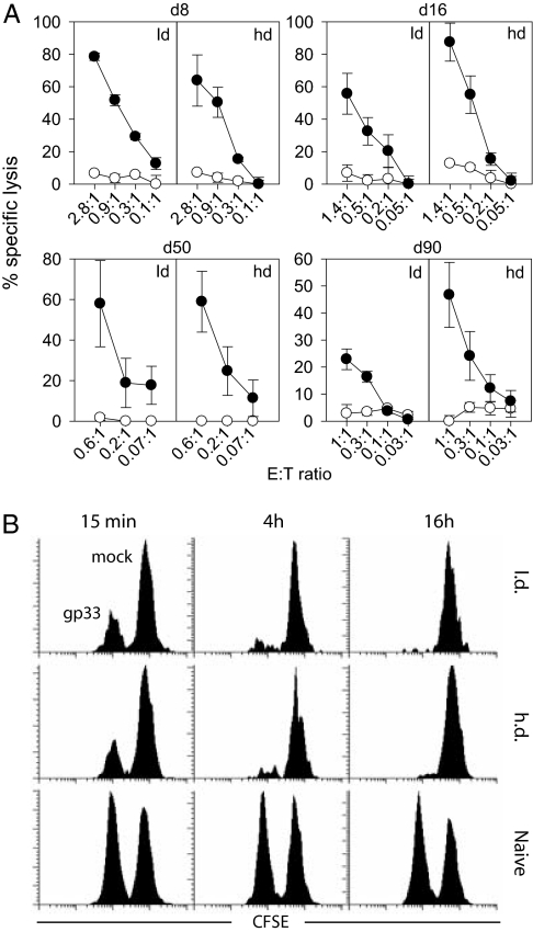
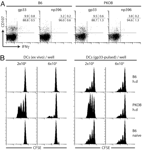
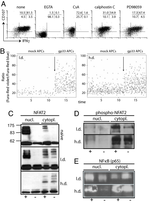
In persistent viral infections, the host's immune system is challenged by the constant exposure to antigen, potentially causing continuous activation of CD8(+) T cells with subsequent immunopathology. Here we demonstrate, for experimental chronic lymphocytic choriomeningitis virus and human HIV infection, that upon prolonged in vivo exposure to antigen, TCR-triggered Ca(2+) flux, degranulation, and cytotoxicity are maintained on a cellular level, whereas cytokine production is severely impaired because of a selective defect in activation-induced NFAT nuclear translocation. During chronic infection, this differential regulation of pathways leading to diverse effector functions may allow CD8(+) T cells to sustain some degree of local viral control by direct cytotoxicity while limiting systemic immune pathology by silencing cytokine production.


Conflict of interest statement

The authors declare no conflict of interest.

Figures

Fig. 1.
Fig. 1.
Functional assessment of CD8+ T cells. (A) C57BL/6 mice were infected i.v. with 200 pfu (l.d.) or 106 pfu (h.d.) LCMV Docile, and 20 days later, gp33- or np396-induced degranulation (CD107) or IFN-γ, TNF-α, and IL-2 production were analyzed for splenocytes. (B) Monoclonal anti-PD-L1 or -CTLA-4 antibodies were added during stimulation. Plots are gated on CD8+ T cells. One of three similar experiments is shown.
Fig. 2.
Fig. 2.
Phenotypical and functional assessment of TCR tg CD8+ T cells. (A) Spleen cells were stained for Ly5.1 and CD43 (gated on CD8+ T cells) or for CD62L and CD127 (gated on CD8+Ly5.1+ T cells). Spleen cells were stimulated with gp33 peptide and degranulation, and IFN-γ production (B) or TNF-α production (C) was assessed. Plots are gated on TCR tg CD8+ T cells (B) or CD8+ T cells (C). (D) Phorbol 12-myristate 13-acetate/ionomycin or gp33 stimulation 16 days after infection. Plots are gated on TCR tg CD8+ T cells.
Fig. 3.
Fig. 3.
Cytotoxic function of LCMV-specific CD8+ T cells. (A) In vitro cytotoxicity. At day 8, 16, 50, or 90 after infection, CD8+ T cells were purified from the spleens of adoptively transfused l.d.- or h.d.-infected mice and adjusted for equal numbers of TCR tg CD8+ T cells for 51Cr release assays (gp33-pulsed: closed circles, mock-pulsed: open circles). Specific lysis was determined after 16 h. Effector-to-target cell ratios indicate the ratio between Ly5.1+ TCR tg CD8+ T cells and target cells. (B) In vivo cytotoxicity. Gp33 peptide-loaded (CFSElo) and unloaded (CFSEhi) target cells were adoptively transferred into infected (day 16) or naïve recipient mice. Target cells were quantified in blood 15 min, 4 h, or 16 h after transfer. Representative plots of three mice per group and of three experiments are shown.
Fig. 4.
Fig. 4.
Chronic LCMV infection in C57BL/6 and PKOB mice. (A) CD8+ T cell function in C57BL/6 and PKOB mice. C57BL/6 and PKOB mice were infected with 106 pfu LCMV Docile, and gp33- and np396-specific CD8+ T cells were analyzed at day 19 in spleen for degranulation and IFN-γ production (gated on CD8+ T cells). (B) Ex vivo quantification of gp33+APCs. CD11c+ DCs were purified to high purity from spleens, were either mock treated (Left) or pulsed with gp33 peptide (Right), serially diluted, and incubated with 2 × 10⁵ purified naïve CFSE-labeled Ly5.1+ TCR tg CD8+ T cells. CFSE dilution profiles were analyzed after 3 days (plots are gated on CD8+ Ly5.1+ cells). One representative plot from three mice and three independent experiments is shown.
Fig. 5.
Fig. 5.
TCR signaling in LCMV-specific CD8+ T cells from h.d. or l.d. infected mice. (A) Signaling requirements for degranulation and IFN-γ production. Spleen cells from adoptively transfused and infected mice (day 20) were stimulated with gp33 peptide in the presence of EGTA, cyclosporine A, calphostin C, or PD98059 and degranulation and IFN-γ production were analyzed. Plots are gated on Ly5.1+ TCR tg CD8+ T cells. One representative staining of two mice and three independent experiments is shown. (B) Ca2+ flux analysis. Spleen cells were isolated, labeled with Fura-red, and stained for Ly5.1 followed by incubation with mock- or gp33-pulsed C57BL/6 peritoneal macrophages; arrows indicate the start of analysis of Ca2+ flux in the presence of gp33-pulsed macrophages. Plots are gated on Ly5.1+ T cells. Representative plots of two mice and three independent experiments are shown. (C–E) Ly5.1 TCR tg CD8+ T cells were sorted from adoptively transfused and h.d.- or l.d.-infected mice or from naïve TCR tg mice. Sorted cells were incubated in presence (+) or absence (−) of gp33 peptide for 16 h. Nuclear and cytoplasmic extracts were prepared and assayed in Western blots for presence of NFAT2 (C; molecular mass, 90, 110, or 140 kDa), p-NFAT2 (D), or NF-κB (E). One of two or three similar experiments is shown.

References

    1. Kaech SM, Ahmed R. Nat Immunol. 2001;2:415–422. - PMC - PubMed
    1. van Stipdonk MJ, Lemmens EE, Schoenberger SP. Nat Immunol. 2001;2:423–429. - PubMed
    1. Kaech SM, Hemby S, Kersh E, Ahmed R. Cell. 2002;111:837–851. - PubMed
    1. Doherty PC, Hou S, Tripp RA. Curr Opin Immunol. 1994;6:545–552. - PubMed
    1. Wherry EJ, Teichgraber V, Becker TC, Masopust D, Kaech SM, Antia R, von Andrian UH, Ahmed R. Nat Immunol. 2003;4:225–234. - PubMed

Publication types