. 2007 Mar 13;104(11):4577-82.
doi: 10.1073/pnas.0700334104. Epub 2007 Mar 5.

Transgenic LacZ under control of Hec-6st regulatory sequences recapitulates endogenous gene expression on high endothelial venules

Affiliations

Transgenic LacZ under control of Hec-6st regulatory sequences recapitulates endogenous gene expression on high endothelial venules

Shan Liao et al. Proc Natl Acad Sci U S A. .

Abstract

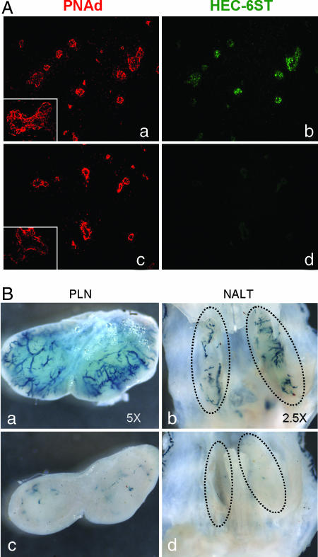
Hec-6st is a highly specific high endothelial venule (HEV) gene that is crucial for regulating lymphocyte homing to lymph nodes (LN). The enzyme is also expressed in HEV-like vessels in tertiary lymphoid organs that form in chronic inflammation in autoimmunity, graft rejection, and microbial infection. Understanding the molecular nature of Hec-6st regulation is crucial for elucidating its function in development and disease. However, studies of HEV are limited because of the difficulties in isolating and maintaining the unique characteristics of these vessels in vitro. The novel pClasper yeast homologous recombination technique was used to isolate from a BAC clone a 60-kb DNA fragment that included the Hec-6st (Chst4) gene with flanking sequences. Transgenic mice were generated with the beta-galactosidase (LacZ) reporter gene inserted in-frame in the exon II of Hec-6st within the isolated BAC DNA fragment. LacZ was expressed specifically on HEV in LN, as indicated by its colocalization with peripheral node vascular addressin. LacZ was increased in nasal-associated lymphoid tissue during development and was reduced in LN and nasal-associated lymphoid tissue by LTbetaR-Ig (lymphotoxin-beta receptor human Ig fusion protein) treatment in a manner identical to the endogenous gene. The transgene was expressed at high levels in lymphoid accumulations with characteristics of tertiary lymphoid organs in the salivary glands of aged mice. Thus, the Hec-6s-LacZ construct faithfully reproduces Hec-6st tissue-specific expression and can be used in further studies to drive expression of reporter or effector genes, which could visualize or inhibit HEV in autoimmunity.


Conflict of interest statement

The authors declare no conflict of interest.

Figures

Fig. 1.
Fig. 1.
Hec-6st transgenic mice. The Hec-6st gene includes two exons, an intron, and flanking sequences. The entire coding sequence is contained in the second exon. The Hec-6st-LacZ transgene construct was generated by isolating from BAC clone 20473A a 60-kb DNA fragment, which included the entire Hec-6st gene with 40-kb upstream and 18-kb downstream noncoding sequence, and inserting in-frame a LacZ cassette composed of the promoterless LacZ reporter gene and URA3 selection marker in yeast into Hec-6st gene exon II by yeast homologous recombination by using pClasper technology. (A) Schematic diagram of the transgene construct. Fg1 and Fg2 were the matching DNA sequences used to insert the LacZ cassette into Hec-6st gene exon II by homologous recombination. FgA and FgB3 were the DNA sequences used to capture the DNA fragments from the BAC clone using the database sequences. URA3A F and Hec-6st R are the PCR primers used for screening the transgenic mice. 5′–3′ represents the database DNA sequence direction. Arrows indicate the direction of gene transcription. (B) Southern blot analysis of tail DNA of Hec-6st-LacZ founder lines. Positive mice are indicated by the presence of a 3.6-kb BamHI fragment hybridizing with a LacZ probe.
Fig. 2.
Fig. 2.
LacZ reproduces HEC-6ST expression on HEV in LN and NALT of the transgenic mice. (A) Transcription of Hec-6st and LacZ mRNA in secondary lymphoid organs (PLN, mesenteric LN, and NALT) as detected by RT-PCR using LacZ and Hec-6st primer pairs reported in Materials and Methods. (B) Expression of LacZ in PLN and NALT analyzed by X-Gal staining for 4 h. (Original magnification: ×2.) (Ba, Bb, and Be) PLN. (Bc and Bd) NALT. (Ba, Bc, and Be) Transgene positive mice. (Bb and Bd) Transgene negative littermates. (Be) LN incubated with X-Gal overnight displays the typical HEV branched pattern. (Original magnification: ×5.) (C) Sections of PLN stained for LacZ (Ca) were costained for PNAd (Cb, red) by immunofluorescence. (Original magnification: ×20.)
Fig. 3.
Fig. 3.
LacZ expression in NALT of transgenic mice mimics wild-type HEC-6ST expression during development. (A) NALT isolated from 3-week-old or 6-week-old C57BL/6 mice. The dotted circles indicate the area of NALT. (Aa and Ac) HE staining. (Original magnification: ×5.) (Ab and Ad) Immunofluorescence double staining for PNAd (red) and HEC-6ST (green). (Original magnification: ×40.) (B) NALT isolated from different lines of Hec-6st-LacZ transgenic mice at 3 weeks or 7–10 weeks of age. (Ba) Line 403, 3 weeks old. (Bb) Line 403, 10 weeks old. (Bc) Line 399, 3 weeks old. (Bd) Line 399, 7 weeks old. LacZ expression was detected by X-Gal staining overnight. (Original magnification: ×2.) Shown are representative results of three experiments of three different Hec-6st-LacZ transgenic mouse lines.
Fig. 4.
Fig. 4.
Diminished expression of HEC-6ST and LacZ in HEV after LTβR-Ig treatment. (A) PLN isolated from C57BL/6 mice 7 days after treatment with LTβR-Ig or control Ig. The expression of HEC-6ST (green) and PNAd (red) in PLN was detected by immunofluorescence double staining. (Aa and Ab) Control Ig-treated mouse. (Ac and Ad) LTβR-Ig-treated mouse. Shown is a representative experiment of n > 3 mice per group. (Original magnification: ×20.) (Aa Inset) Typical HEV with luminal and abluminal PNAd staining. (Ac Inset) Typical HEV with abluminal PNAd staining after LTβR-Ig treatment. (Original magnification of Insets: ×100.) (B) PLN and NALT isolated at day 7 after treatment with LTβR-Ig or control Ig. LacZ expression was detected by X-Gal staining overnight. (Ba and Bb) Control Ig-treated mouse. (Bc and Bd) LTβR-Ig-treated mouse. Shown are representative experiments of n = 3 mice per group.
Fig. 5.
Fig. 5.
HEC-6ST and LacZ expression on HEV in spontaneous TLO in salivary glands. (A–D) Absence of TLO in salivary glands isolated from a 9-month-old transgene positive mouse. (A) H&E-stained section. (B) CD3 (red) and B220 (green) double immunofluorescence stained frozen section. (C) PNAd (red) and lymphatic vessel endothelial hyaluronan receptor 1 (green) double immunofluorescence and HE-counterstained frozen section. (D) X-Gal staining of the entire salivary gland. (Original magnification: ×20 in A–C and ×2 in D.) (E–J) TLO in salivary glands isolated from a 15-month-old transgene-positive mouse (a sibling of the above mouse). (E) H&E-stained section. (F) CD3 (red) and B220 (green) double immunofluorescence stained frozen section. (G) PNAd (red) and lymphatic vessel endothelial hyaluronan receptor 1 (green) double immunofluorescence and HE-counterstained frozen section. Note that the lymphatic sinus is packed with lymphocytes. (Original magnification: ×20 in E–G.) (H) X-Gal staining of the entire salivary gland. (Original magnification: ×2.) (I) HE-counterstaining of a 7-μm section of the X-Gal-stained salivary gland reveals that LacZ is expressed exclusively within the TLO. (J) The X-Gal-stained salivary glands were sectioned and stained for PNAd. LacZ (blue) and PNAd (red) are colocalized in one TLO of the salivary gland. (Original magnification: ×40 in I and J.)

Similar articles

Cited by

References

    1. Berg EL, Robinson MK, Warnock RA, Butcher EC. J Cell Biol. 1991;114:343–349. - PMC - PubMed
    1. Streeter PR, Berg EL, Rouse BT, Bargatze RF, Butcher EC. Nature. 1988;331:41–46. - PubMed
    1. Hemmerich S, Rosen SD. Glycobiology. 2000;10:849–856. - PubMed
    1. Shailubhai K, Streeter PR, Smith CE, Jacob GS. Glycobiology. 1997;7:305–314. - PubMed
    1. Hemmerich S, Butcher EC, Rosen SD. J Exp Med. 1994;180:2219–2226. - PMC - PubMed

Publication types

LinkOut - more resources