. 2007 Mar 13;104(11):4588-93.
doi: 10.1073/pnas.0611653104. Epub 2007 Mar 7.

Impaired angiogenesis in aminopeptidase N-null mice

Affiliations

Impaired angiogenesis in aminopeptidase N-null mice

Roberto Rangel et al. Proc Natl Acad Sci U S A. .

Abstract

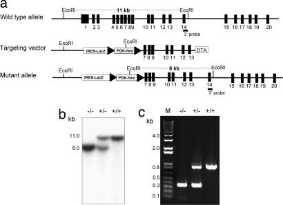
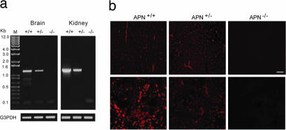
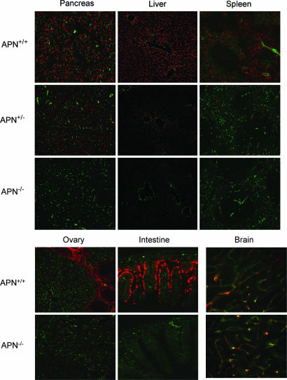
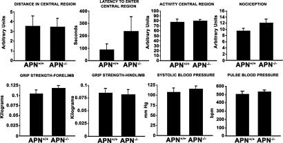
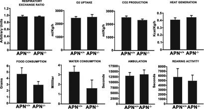
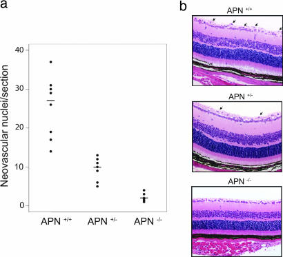
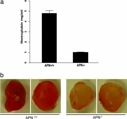
Aminopeptidase N (APN, CD13; EC 3.4.11.2) is a transmembrane metalloprotease with several functions, depending on the cell type and tissue environment. In tumor vasculature, APN is overexpressed in the endothelium and promotes angiogenesis. However, there have been no reports of in vivo inactivation of the APN gene to validate these findings. Here we evaluated, by targeted disruption of the APN gene, whether APN participates in blood vessel formation and function under normal conditions. Surprisingly, APN-null mice developed with no gross or histological abnormalities. Standard neurological, cardiovascular, metabolic, locomotor, and hematological studies revealed no alterations. Nonetheless, in oxygen-induced retinopathy experiments, APN-deficient mice had a marked and dose-dependent deficiency of the expected retinal neovascularization. Moreover, gelfoams embedded with growth factors failed to induce functional blood vessel formation in APN-null mice. These findings establish that APN-null mice develop normally without physiological alterations and can undergo physiological angiogenesis but show a severely impaired angiogenic response under pathological conditions. Finally, in addition to vascular biology research, APN-null mice may be useful reagents in other medical fields such as malignant, cardiovascular, immunological, or infectious diseases.


Conflict of interest statement

The authors declare no conflict of interest.

Figures

Fig. 1.
Fig. 1.
Generation and characterization of the APN-null mouse. (a) Schema of the APN gene-targeting strategy. The APN gene contains 20 exons (black boxes); the 3′ external probe is located in exon 14. The targeting vector contains a β-galactosidase transgene and a floxed neomycin resistant gene. (b) Southern blot of genomic DNA from WT (+/+), APN-heterozygous (+/−), and APN-null (−/−); the mutant allele (8 kb) and the WT allele (11 kb) are noted. The APN heterozygous contains both alleles. (c) PCR genotyping reveals unique products for WT (879-bp) and APN-null (320-bp) mice. The APN heterozygous contains both PCR products.
Fig. 2.
Fig. 2.
Expression analysis of the APN-null mouse. (a) RT-PCR analysis of total RNA. The WT (+/+) and heterozygous (+/−) mice contain the 1.2-kb PCR product representing the APN mRNA transcript that is undetectable in the APN-null mouse (−/−). The G3PDH transcript served as a loading control. (b) Immunohistochemical analysis of the APN protein in brain (Upper) and kidney (Lower). APN immunoreactivity (red fluorescence) is detected in renal tubules and in pericytes within brain vasculature of WT and heterozygous mice but not in the APN-null mice (Right). (Scale bar, 100 μm.)
Fig. 3.
Fig. 3.
Immunohistochemical analysis of APN and CD31. Colocalization studies in pancreas, liver, and spleen for APN (red fluorescence) and CD31 (green fluorescence) were performed in frozen tissue sections (Upper). The WT and APN-heterozygote mice contain dual fluorescence signals; in APN-null mice, only CD31 immunoreactivity is detected. Ovary and intestine were also evaluated (Lower Left). Brain was costained with anti-CD31 and -APA antibodies. The yellow fluorescence reflects the localization of both proteins in pericytes in brain vasculature (Lower Right).
Fig. 4.
Fig. 4.
Noninvasive phenotypic studies. Spontaneous locomotor activity in WT mice and APN-null littermates are represented as distance in central region, latency central region, and activity central region. Nociception was measured by using a hot-plate analgesia meter. Neuromuscular function was determined by grip strength of the forelimb and hindlimb. Pulse and blood pressure were measured by the tail-cuff method. Error bars indicate SEM of WT and APN-null mice (n = 6 per group).
Fig. 5.
Fig. 5.
Comprehensive in-cage monitoring system. The WT and APN-null mice littermates were examined for respiratory exchange ratio, oxygen uptake, and carbon dioxide production. Caloric intake and energy metabolism were measured for heat production, food, and water consumption. Ambulation and rearing activity were determined by using the comprehensive laboratory animal-monitoring system. Error bars indicate SEM of WT and APN-null mice (n = 6 per group).
Fig. 6.
Fig. 6.
Failed retinal neovascularization in APN-null mice under hypoxic conditions. (a) Quantification of endothelial cells nuclei extended across the inner surface of the retina into the vitreous space. Data from WT, APN-heterozygous, and APN-null mice were compared (P < 0.001). (b) APN-null mice lack the nuclei protruding into the vitreous space of the eye, as observed in the WT and heterozygote representative examples (arrows). Five independent experiments were performed with similar results.
Fig. 7.
Fig. 7.
APN-null mice have severely impaired angiogenesis upon growth factor stimulation. (a) Hemoglobin content of the gelfoam from WT and APN-null mice were analyzed. (b) Gelfoam plugs implanted in the s.c. tissue were removed after 14 days and analyzed. Plugs in WT mice contained significantly more hemoglobin (4.8 ± 0.3 mg/ml) than those in APN−/− mice (1.0 ± 0.1 mg/ml; P < 0.0001). Five independent experiments were performed with similar results.

Similar articles

Cited by

References

    1. Hooper NM, Lendeckel U, editors. Aminopeptidases in Biology and Disease. New York: Kluwer Academic/Plenum; 2004.
    1. Barrett AJ, Rawlings ND, Woessner JF, editors. Handbook of Proteolytic Enzymes. London: Elsevier; 2004.
    1. Amoscato AA, Alexander JW, Babcock GF. J Immunol. 1989;142:1245–1252. - PubMed
    1. Favaloro EJ, Bradstock KF, Kabral A, Grimsley P, Zowtyj H, Zola H. Br J Haematol. 1988;69:163–171. - PubMed
    1. Mechtersheimer G, Moller P. Am J Pathol. 1990;137:1215–1222. - PMC - PubMed

Publication types