. 2007 Mar 13;104(11):4664-9.
doi: 10.1073/pnas.0603243104. Epub 2007 Mar 5.

Pleiotrophin is a neurotrophic factor for spinal motor neurons

Affiliations

Pleiotrophin is a neurotrophic factor for spinal motor neurons

Ruifa Mi et al. Proc Natl Acad Sci U S A. .

Abstract

Regeneration in the peripheral nervous system is poor after chronic denervation. Denervated Schwann cells act as a "transient target" by secreting growth factors to promote regeneration of axons but lose this ability with chronic denervation. We discovered that the mRNA for pleiotrophin (PTN) was highly up-regulated in acutely denervated distal sciatic nerves, but high levels of PTN mRNA were not maintained in chronically denervated nerves. PTN protected spinal motor neurons against chronic excitotoxic injury and caused increased outgrowth of motor axons out of the spinal cord explants and formation of "miniventral rootlets." In neonatal mice, PTN protected the facial motor neurons against cell death induced by deprivation from target-derived growth factors. Similarly, PTN significantly enhanced regeneration of myelinated axons across a graft in the transected sciatic nerve of adult rats. Our findings suggest a neurotrophic role for PTN that may lead to previously unrecognized treatment options for motor neuron disease and motor axonal regeneration.


Conflict of interest statement

The authors declare no conflict of interest.

Figures

Fig. 1.
Fig. 1.
Changes in PTN expression. (A) PTN mRNA expression was measured by using quantitative RT-PCR in rat sciatic nerves after transection at the midthigh level. PTN mRNA was up-regulated in denervated distal nerves, but this up-regulation was not maintained (n = 4–6 animals per time point; ∗, P < 0.005 compared with contralateral intact side). (B) Expression of PTN mRNA in rat tibialis anterior muscle during development and after denervation by transection of the sciatic nerve at the midthigh level (n = 4 animals per time point; ∗, P < 0.005 compared with levels of expression in normal adult muscle).
Fig. 2.
Fig. 2.
PTN is neurotrophic for spinal motor neurons. (A) Neurotrophism of PTN was examined in spinal cord explants prepared from postnatal-day-8 rats. The explants were cultured on semipermeable membrane inserts with a point source of PTN in a gelfoam away from the explants, and after 1 week the cultures were stained with antineurofilament antibody SMI-32. The motor neurons were identified by their size and location within the explant. PTN induced axonal outgrowth from spinal cord explants and formation of miniventral rootlets toward the source of PTN; a representative explant is shown. In cultures without PTN, axons remained at the gray–white matter junction (arrow) and never exited the explants. The images are representative of n = 12–16 per group. (Scale bar, 150 μm.) (B) In cultures treated with a diffuse source of PTN in the culture medium, PTN increased the number of spinal motor axons that crossed the gray–white matter junction and entered the white matter tracts. (Scale bar, 30 μm.) (C) Quantitation was done by counting the number of axons crossing the gray–white matter junction in the ventral half of the spinal cord explants (n = 12 explants per group; ∗, P < 0.005).
Fig. 3.
Fig. 3.
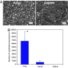
PTN promotes regeneration of axons after sciatic nerve transection. (A) In adult rats, sciatic nerves were transected at the midthigh level and repaired with silicone tubes filled with HEK-293PTN cells, HEK-293vector cells, or saline. Images are from 1-μm transverse sections embedded in plastic at 12 mm distal to the proximal repair site. There were many myelinated axons in the animals treated with HEK-293PTN cells (Left) compared with the animals treated with HEK-293vector cells (Right). There were no regenerated axons in the saline-treated animals. (B) Quantitation of myelinated fibers shows that more axons regenerated into the silicone tubes filled with HEK-293PTN cells compared with HEK-293vector or saline-filled tubes (n = 8–9 per group; ∗, P < 0.005 compared with the other two groups).
Fig. 4.
Fig. 4.
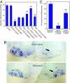
PTN is neuroprotective. (A) Spinal cord explant cultures were treated with glutamate transport inhibitor, THA, with or without PTN for 4 weeks. Then explants were stained with antineurofilament antibody, and motor neurons in the ventral half of the explants were counted. PTN protected spinal motor neurons against chronic excitotoxicity induced by glutamate transport inhibition (n = 8 per condition; ∗, P < 0.005 compared with control; ∗∗, P < 0.005 compared with THA alone). (B) Three-day-old mouse pups had a transection of the facial nerve on one side and treated with gelfoams loaded with HEK-293PTN or HEK-293vector at the stump of the nerves. A week later, cryostat-sectioned brainstems were stained with cresyl violet. Arrows indicate the transected side. (C) Quantitation of the facial motor nucleus neuron counts showed that PTN protected neonatal facial motor neurons against cell death induced by facial nerve transection (contralat., contralateral; ipsilat., ipsilateral; n = 6 per condition; ∗, P < 0.005 compared with control; ∗∗, P < 0.005 compared with vector).
Fig. 5.
Fig. 5.
ALK mediates neurotrophic activity of PTN. (A) Changes in mRNA levels of PTN receptors in the adult rat ventral spinal cord and DRG (L4 and L5 levels) were evaluated by quantitative RT-PCR 1 week after sciatic nerve transection. Values are expressed as percentages of change from ventral spinal cords and DRGs with intact sciatic nerves (∗, P < 0.005). (B) Spinal cord explants from postnatal day 8 were double-stained with antineurofilament antibody (NF) and anti-ALK antibody (ALK); a spinal motor neuron is shown. (C) Spinal cord explants were treated with conditioned media (CM) from HEK-293vector or HEK-293PTN with and without blocking anti-ALK antibody or a control antibody (anti-GFAP) for 3 days. Then cultures were stained with antineurofilament antibody, and the number of axons crossing the gray–white matter junction in the ventral half and exiting from the explant was counted (n = 8 per condition; ∗, P < 0.005 compared with HEK-293vector conditioned media; ∗∗, P < 0.005 compared with HEK-293PTN and HEK-293PTN plus anti-GFAP antibody). (D) Spinal cord explants were treated with PTN, THA, anti-ALK, or anti-GFAP antibodies for 4 weeks. Surviving motor neuron numbers were counted (n = 10 per condition; ∗, P < 0.005 compared with control; ∗∗, P < 0.05 compared with THA alone; ∗∗∗, P < 0.05 compared with THA plus PTN or THA plus PTN plus anti-ALK).

Similar articles

Cited by

References

    1. Boyd JG, Gordon T. Exp Neurol. 2003;183:610–619. - PubMed
    1. Frostick SP, Yin Q, Kemp GJ. Microsurgery. 1998;18:397–405. - PubMed
    1. Fu SY, Gordon T. J Neurosci. 1995;15:3886–3895. - PMC - PubMed
    1. Sulaiman OA, Gordon T. Glia. 2000;32:234–246. - PubMed
    1. Sunderland S. Nerves and Nerve Injuries. Edinburgh: Livingstone; 1978.

Publication types