. 2007 Mar 13;104(11):4688-93.
doi: 10.1073/pnas.0606067104. Epub 2007 Mar 5.

Mineralocorticoid receptor overexpression in forebrain decreases anxiety-like behavior and alters the stress response in mice

Affiliations

Mineralocorticoid receptor overexpression in forebrain decreases anxiety-like behavior and alters the stress response in mice

Aaron M Rozeboom et al. Proc Natl Acad Sci U S A. .

Abstract

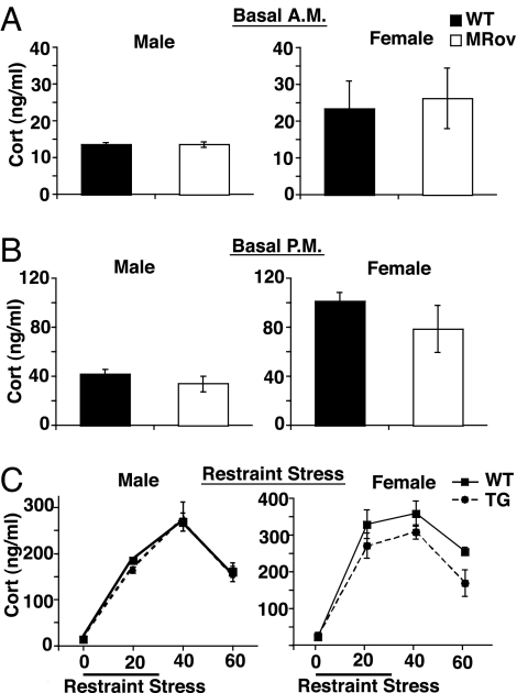
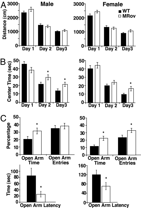
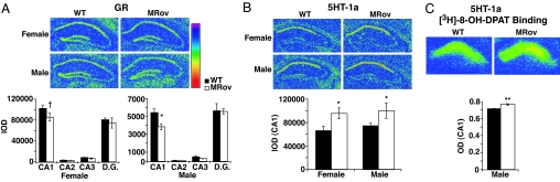
Although numerous stress-related molecules have been implicated in vulnerability to psychiatric illness, especially major depression and anxiety disorders, the role of the brain mineralocorticoid receptor (MR) in stress, depression, and affective function is not well defined. MR is a steroid hormone receptor that detects circulating glucocorticoids with high affinity and has been primarily implicated in controlling their basal level and circadian rhythm. To specifically address the role of MR in hypothalamic-pituitary-adrenal axis activity and anxiety-related behaviors, we generated transgenic mice with increased levels of MR in the forebrain (MRov mice) by using the forebrain-specific calcium/calmodulin-dependent protein kinase II alpha promoter to direct expression of MR cDNA. A mild but chronic elevation in forebrain MR results in decreased anxiety-like behavior in both male and female transgenic mice. Female MRov mice also exhibit a moderate suppression of the corticosterone response to restraint stress. Increased forebrain MR expression alters the expression of two genes associated with stress and anxiety, leading to a decrease in the hippocampal glucocorticoid receptor (GR) and an increase in serotonin receptor 5HT-1a, consistent with the decreased anxiety phenotype. These data suggest that the functions of forebrain MR may overlap with GR in hypothalamic-pituitary-adrenal axis regulation, but they dissociate significantly from GR in the modulation of affective responses, with GR overexpression increasing anxiety-like behavior and MR overexpression dampening it. These findings point to the importance of the MR:GR ratio in the control of emotional reactivity.


Conflict of interest statement

The authors declare no conflict of interest.

Figures

Fig. 1.
Fig. 1.
Transgene construct and properties of Flag-MR. (A) Schematic of transgene construct. The CaMKIIα promoter directs expression of a 3×Flag-tagged mouse MR cDNA. Arrows represent the location of the primer pair used for PCR genotyping. The two bars represent the locations of the two probes used for ISH analysis (TG, transgene specific; MR, endogenous MR + transgene directed MR). NTD, N-terminal domain; DBD, DNA-binding domain; LBD, ligand-binding domain. (B) Transactivation of glucocorticoid responsive element (GRE)-luciferase by MR vs. Flag-MR. CV-1 cells were transfected with MR, Flag-MR or vector alone, GRE-luciferase, and CMV-βgal; 24 h later, cells were treated with drugs for an additional 24 h (corticosterone = 100 nM, aldosterone = 10 nM). Normalized luciferase activity (luciferase activity/βgal activity) for samples was divided by their respective no drug control to result in fold induction of reporter gene activity. The data are presented as mean ± SEM and represent three to five independent experiments done in duplicate. (C) Ligand-binding properties of MR vs. Flag-MR. Cos-1 cells were transfected with MR or Flag-MR. Cells were harvested after 40 h, and [3H]corticosterone was used to determine dissociation constants (Kd) for MR and Flag-MR in five independent Scatchard analyses. Data represent mean ± SEM. Experimental details are in SI Methods.
Fig. 2.
Fig. 2.
Forebrain-specific transgene expression in MRov mice. (A) ISH with MR and transgene-specific (TG) probes on sagittal sections from wild type (WT) and two different MRov founder lines reveals forebrain specificity of transgene expression. (B) Western blot analysis with anti-Flag antibody confirms expression of Flag-MR in forebrain regions of both male (Upper) and female (Lower) mice. Regions include hippocampus (Hip), hypothalamus (Hyp), cortex (Ctx), hindbrain/cerebellum (HB), and pituitary (Pit). (C) Representative pseudocolored ISH images with an MR-specific cRNA probe show overexpression of MR mRNA in subregions of the hippocampus (blue = low expression, red = high expression). Quantitation represents integrated optical density (IOD) analysis of 4–5 animals/genotype/sex and 9–14 sections/animal. Data are presented as mean ± SEM. ∗, P < 0.05 by t test.
Fig. 3.
Fig. 3.
Normal basal HPA axis activity, but sexually dimorphic suppression of corticosterone release in response to restraint stress. (A) Basal a.m. corticosterone levels (n = 4–5/genotype/sex). (B) basal p.m. corticosterone levels (n = 5–6/genotype/sex). (C) Restraint stress-induced corticosterone release in male and female MRov (TG) and wild-type (WT) mice in the a.m. Female MRov mice exhibit a moderate suppression of the corticosterone response to stress over the stress and recovery time course. ANOVA reveals a significant difference due to genotype (P = 0.029, n = 4–10/genotype/time point).
Fig. 4.
Fig. 4.
Reduced anxiety-like behavior in forebrain MRov mice. (A) Three days of open field exposure show normal habituation and no changes in locomotor activity in either male or female MRov mice (first 6 min of activity shown). (B–C) Anxiety-like behavior measures in male and female MRov mice. (B) Open field activity reveals increased center time on days 2 and/or 3. (C) MRov mice show decreases in measures of anxiety-like behavior on the EPM, with significant changes in percentage of open arm time, percentage of open arm entries (females only), and open arm latency. Data represent mean ± SEM of 19–40 animals/genotype/sex. ∗, P < 0.05.
Fig. 5.
Fig. 5.
Altered expression of GR and 5HT-1a in MRov mice. (A) ISH analyses with a GR-specific probe show a decrease in GR mRNA in region CA1 of the hippocampus of male MRov mice and a trend toward decreased GR in region CA1 of female MRov mice (∗, P < 0.05, †, P = 0.11) (n = 5 animals/genotype/sex, and 12–16 sections/animal for males and 7–9 sections/animal for females). (B) ISH with a 5HT-1a-specific probe shows significantly increased levels of the receptor mRNA in the CA1 of male and female MRov mice (n = 4–5 animals/genotype/sex, and 9–12 sections/animal). (∗, P < 0.05.) (C) 5HT-1a receptor binding with [3H]8-OH-DPAT shows significantly increased levels of binding (∗∗, P = 0.0004) (n = 6 males/genotype and 8–10 sections/animal). Representative pseudocolored images are shown.

References

    1. Chrousos GP, Gold PW. J Am Med Assoc. 1992;267:1244–1252. - PubMed
    1. De Kloet ER, Vreugdenhil E, Oitzl MS, Joels M. Endocr Rev. 1998;19:269–301. - PubMed
    1. Akil H. Nature Medicine. 2005;11:116–118. - PubMed
    1. Lopez JF, Chalmers DT, Little KY, Watson SJ. Biol Psychiatry. 1998;43:547–573. - PubMed
    1. Patel PD, Lopez JF, Lyons DM, Burke S, Wallace M, Schatzberg AF. J Psychiatr Res. 2000;34:383–392. - PubMed

Publication types

Substances