

Structure of the non-redox-active tungsten/[4Fe:4S] enzyme acetylene hydratase
- PMID: 17360611
- PMCID: PMC1805521
- DOI: 10.1073/pnas.0610407104
Structure of the non-redox-active tungsten/[4Fe:4S] enzyme acetylene hydratase
Abstract
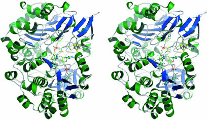
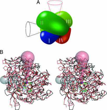
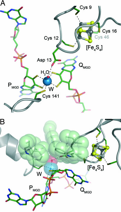
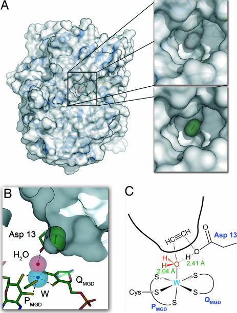
The tungsten-iron-sulfur enzyme acetylene hydratase stands out from its class because it catalyzes a nonredox reaction, the hydration of acetylene to acetaldehyde. Sequence comparisons group the protein into the dimethyl sulfoxide reductase family, and it contains a bis-molybdopterin guanine dinucleotide-ligated tungsten atom and a cubane-type [4Fe:4S] cluster. The crystal structure of acetylene hydratase at 1.26 A now shows that the tungsten center binds a water molecule that is activated by an adjacent aspartate residue, enabling it to attack acetylene bound in a distinct, hydrophobic pocket. This mechanism requires a strong shift of pK(a) of the aspartate, caused by a nearby low-potential [4Fe:4S] cluster. To access this previously unrecognized W-Asp active site, the protein evolved a new substrate channel distant from where it is found in other molybdenum and tungsten enzymes.
Conflict of interest statement
The authors declare no conflict of interest.
Figures
References
-
- Fráusto da Silva JJR, Williams RJP. The Biological Chemistry of the Elements: The Inorganic Chemistry of Life. Oxford, UK: Oxford Univ Press; 2001.
-
- Dobbek H, Huber R. Molybdenum and Tungsten: Their Roles in Biological Processes. Vol 39. 2002. pp. 227–263. - PubMed
-
- Kisker C, Schindelin H, Baas D, Retey J, Meckenstock RU, Kroneck PMH. FEMS Microbiol Rev. 1998;22:503–521. - PubMed
-
- Bertero MG, Rothery RA, Palak M, Hou C, Lim D, Blasco F, Weiner JH, Strynadka NCJ. Nat Struct Biol. 2003;10:681–687. - PubMed
-
- Dias JM, Than ME, Humm A, Huber R, Bourenkov GP, Bartunik HD, Bursakov S, Calvete J, Caldeira J, Carneiro C, et al. Structure. 1999;7:65–79. - PubMed
Publication types
MeSH terms
Substances
Associated data
- Actions
LinkOut - more resources
Full Text Sources
Other Literature Sources
Molecular Biology Databases
Miscellaneous
