

Evidence for a dual binding mode of dockerin modules to cohesins
- PMID: 17360613
- PMCID: PMC1805526
- DOI: 10.1073/pnas.0611173104
Evidence for a dual binding mode of dockerin modules to cohesins
Abstract
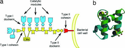
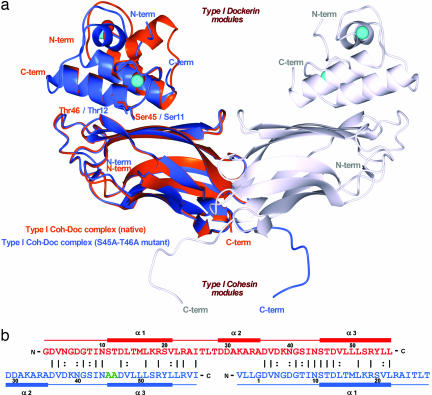
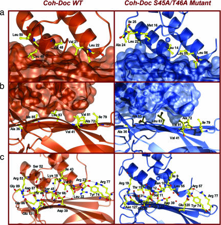
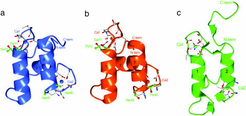
The assembly of proteins that display complementary activities into macromolecular complexes is critical to cellular function. One such enzyme complex, of environmental significance, is the plant cell wall degrading apparatus of anaerobic bacteria, termed the cellulosome. The complex assembles through the interaction of enzyme-derived "type I dockerin" modules with the multiple "cohesin" modules of the scaffolding protein. Clostridium thermocellum type I dockerin modules contain a duplicated 22-residue sequence that comprises helix-1 and helix-3, respectively. The crystal structure of a C. thermocellum type I cohesin-dockerin complex showed that cohesin recognition was predominantly through helix-3 of the dockerin. The sequence duplication is reflected in near-perfect 2-fold structural symmetry, suggesting that both repeats could interact with cohesins by a common mechanism in wild-type (WT) proteins. Here, a helix-3 disrupted mutant dockerin is used to visualize the reverse binding in which the dockerin mutant is indeed rotated 180 degrees relative to the WT dockerin such that helix-1 now dominates recognition of its protein partner. The dual binding mode is predicted to impart significant plasticity into the orientation of the catalytic subunits within this supramolecular assembly, which reflects the challenges presented by the degradation of a heterogeneous, recalcitrant, insoluble substrate by a tethered macromolecular complex.
Conflict of interest statement
The authors declare no conflict of interest.
Figures
Similar articles
-
Novel Clostridium thermocellum type I cohesin-dockerin complexes reveal a single binding mode.J Biol Chem. 2012 Dec 28;287(53):44394-405. doi: 10.1074/jbc.M112.407700. Epub 2012 Nov 1. J Biol Chem. 2012. PMID: 23118225 Free PMC article.
-
Structure-function analyses generate novel specificities to assemble the components of multienzyme bacterial cellulosome complexes.J Biol Chem. 2018 Mar 16;293(11):4201-4212. doi: 10.1074/jbc.RA117.001241. Epub 2018 Jan 24. J Biol Chem. 2018. PMID: 29367338 Free PMC article.
-
The Clostridium cellulolyticum dockerin displays a dual binding mode for its cohesin partner.J Biol Chem. 2008 Jun 27;283(26):18422-30. doi: 10.1074/jbc.M801533200. Epub 2008 Apr 28. J Biol Chem. 2008. PMID: 18445585
-
Cellulosomes: microbial nanomachines that display plasticity in quaternary structure.Mol Microbiol. 2007 Mar;63(6):1568-76. doi: 10.1111/j.1365-2958.2007.05640.x. Mol Microbiol. 2007. PMID: 17367380 Review.
-
Single versus dual-binding conformations in cellulosomal cohesin-dockerin complexes.Curr Opin Struct Biol. 2016 Oct;40:89-96. doi: 10.1016/j.sbi.2016.08.002. Epub 2016 Aug 28. Curr Opin Struct Biol. 2016. PMID: 27579515 Review.
Cited by
-
Purification, crystallization and preliminary X-ray characterization of the third ScaB cohesin in complex with an ScaA X-dockerin from Acetivibrio cellulolyticus.Acta Crystallogr F Struct Biol Commun. 2014 May;70(Pt 5):656-8. doi: 10.1107/S2053230X1400750X. Epub 2014 Apr 25. Acta Crystallogr F Struct Biol Commun. 2014. PMID: 24817731 Free PMC article.
-
Complexity of the Ruminococcus flavefaciens FD-1 cellulosome reflects an expansion of family-related protein-protein interactions.Sci Rep. 2017 Feb 10;7:42355. doi: 10.1038/srep42355. Sci Rep. 2017. PMID: 28186207 Free PMC article.
-
The Electrosome: A Surface-Displayed Enzymatic Cascade in a Biofuel Cell's Anode and a High-Density Surface-Displayed Biocathodic Enzyme.Nanomaterials (Basel). 2017 Jun 23;7(7):153. doi: 10.3390/nano7070153. Nanomaterials (Basel). 2017. PMID: 28644390 Free PMC article.
-
Lignocellulose degradation in bacteria and fungi: cellulosomes and industrial relevance.Front Microbiol. 2025 Apr 25;16:1583746. doi: 10.3389/fmicb.2025.1583746. eCollection 2025. Front Microbiol. 2025. PMID: 40351319 Free PMC article. Review.
-
Small angle X-ray scattering analysis of Clostridium thermocellum cellulosome N-terminal complexes reveals a highly dynamic structure.J Biol Chem. 2013 Mar 15;288(11):7978-7985. doi: 10.1074/jbc.M112.408757. Epub 2013 Jan 22. J Biol Chem. 2013. PMID: 23341454 Free PMC article.
References
-
- Warren RA. Annu Rev Microbiol. 1996;50:183–212. - PubMed
-
- Himmel ME, Ruth MF, Wyman CE. Curr Opin Biotechnol. 1999;10:358–364. - PubMed
-
- Bhat MK. Biotechnol Adv. 2000;18:355–383. - PubMed
-
- Boudet AM, Kajita S, Grima-Pettenati J, Goffner D. Trends Plants Sci. 2003;8:576–581. - PubMed
-
- Ragauskas AJ, Williams CK, Davison BH, Britovsek G, Cairney J, Eckert CA, Frederick WJ, Jr, Hallett JP, Leak DJ, Liotta CL, et al. Science. 2006;11:484–489. - PubMed
Publication types
MeSH terms
Substances
Associated data
- Actions
LinkOut - more resources
Full Text Sources
Other Literature Sources
Molecular Biology Databases
