. 2007 Feb 27;104(9):3113-8.
doi: 10.1073/pnas.0608725104. Epub 2007 Feb 20.

Structural basis and evolutionary origin of actin filament capping by twinfilin

Affiliations

Structural basis and evolutionary origin of actin filament capping by twinfilin

Ville O Paavilainen et al. Proc Natl Acad Sci U S A. .

Abstract

Dynamic reorganization of the actin cytoskeleton is essential for motile and morphological processes in all eukaryotic cells. One highly conserved protein that regulates actin dynamics is twinfilin, which both sequesters actin monomers and caps actin filament barbed ends. Twinfilin is composed of two ADF/cofilin-like domains, Twf-N and Twf-C. Here, we reveal by systematic domain-swapping/inactivation analysis that the two functional ADF-H domains of twinfilin are required for barbed-end capping and that Twf-C plays a critical role in this process. However, these domains are not functionally equivalent. NMR-structure and mutagenesis analyses, together with biochemical and motility assays showed that Twf-C, in addition to its binding to G-actin, interacts with the sides of actin filaments like ADF/cofilins, whereas Twf-N binds only G-actin. Our results indicate that during filament barbed-end capping, Twf-N interacts with the terminal actin subunit, whereas Twf-C binds between two adjacent subunits at the side of the filament. Thus, the domain requirement for actin filament capping by twinfilin is remarkably similar to that of gelsolin family proteins, suggesting the existence of a general barbed-end capping mechanism. Furthermore, we demonstrate that a synthetic protein consisting of duplicated ADF/cofilin domains caps actin filament barbed ends, providing evidence that the barbed-end capping activity of twinfilin arose through a duplication of an ancient ADF/cofilin-like domain.


Conflict of interest statement

The authors declare no conflict of interest.

Figures
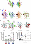
Fig. 1.
Fig. 1.
Twinfilin's C-terminal ADF-H domain is structurally and functionally homologous to ADF/cofilin. (A) Schematic ribbon representation of the overall fold of Twf-C at two orientations rotated by 90°. The sequence is color coded from N to C terminus with blue through yellow to red. (B) A Cα-superimposition of Twf-C with Twf-N (28) (PDB ID code 1M4J) and yeast cofilin (32) (PDB ID code 1COF) and Twf-N with gelsolin segment-1 (PDB ID code 1D0N) and yeast cofilin. The β-strands 3 and 4 are oriented differently in Twf-N (red arrow) and Twf-C (blue arrow). (C) Ribbon diagrams of twinfilin1–142, twinfilin176–316, yeast cofilin, and gelsolin segment 1. The side chains of the residues important for actin-monomer binding are indicated in red. The side chains important for actin-filament binding are indicated in blue, and the Twf-C residues mutated in this study that do not contribute to actin-monomer binding are indicated in green. The Twf-C residues mutated in this study are indicated by letters and numbers (see SI Fig. 6 for the data). The residues important for actin binding in Twf-N cofilin and gelsolin segment 1 are from refs. , , and . (D) The amount of Twf-N or Twf-C (5 μM) cosedimenting with 10 μM actin filaments was determined. Twf-N does not shift to the pellet fraction upon addition of F-actin, whereas Twf-C cosediments with F-actin. The C-terminal tail region increases the affinity of Twf-C for F-actin. (E) Actin filament pointed-end depolymerization assay. 3 μM gelsolin-capped 50% pyrene-labeled actin filaments were diluted to 0.11 μM, and the rate of depolymerization was plotted at different twinfilin domain concentrations. Twinfilin's N-terminal ADF-H domain does not induce filament depolymerization, whereas the construct containing twinfilin's C-terminal ADF-H domain and the tail region increases the pointed-end depolymerization rate by ≈15-fold. Twinfilin's C-terminal ADF-H domain without the tail region increases pointed-end depolymerization by ≈2.5-fold (Inset).
Fig. 2.
Fig. 2.
Interaction of twinfilin domain-swap mutants with G-actin and filament barbed ends. (A) The change in the fluorescence of NBD-labeled Mg-ADP–G-actin was measured at different concentrations of the mutant proteins. Symbols indicate data, and solid lines indicate fitted binding curves for a complex with 1:1 stoichiometry (1:2 stoichiometry for the C-C mutant). (B) The F-actin capping activity of the domain-swap mutants measured by a pyrene–actin polymerization assay. Barbed-end growth (filled circles) and pointed-end growth (open squares) were initiated by using spectrin–actin seeds and gelsolin–actin seeds, respectively, in the presence of 2.5 μM G-actin (10% pyrenyl-labeled) and twinfilin as indicated. The initial rates were normalized to the value of 1 measured in the absence of twinfilin. Similarly to wild-type twinfilin (23) the inhibition of barbed-end polymerization was stronger at low concentrations of C-C, C-N, and N-C constructs than expected from monomer sequestering, indicating barbed-end capping. In contrast, the protein composed of two N-terminal domains did not cap barbed ends based on this assay. The obtained KT (ATP–G-actin sequestration) and KF values (barbed-end capping) are depicted in the figure. (C) The abilities of the mutant proteins to replace capping protein in a biomimetic motility assay were tested. Wild-type twinfilin as well as C-C, C-N, and N-C constructs rescued the tail morphology and bead motility. Also the N-N mutant was partially capable in rescuing bead motility, although the morphology of actin tails was severely abnormal in the presence of this mutant.
Fig. 3.
Fig. 3.
A hybrid protein composed of two ADF/cofilins fused together by twinfilin's linker region caps actin-filament barbed ends. (A) The effects of 0 μM or 0.2 μM cofilin-2, and 0.2 μM cof-cof mutant on the bead motility in the absence of barbed-end capper. Addition of the cof-cof mutant, but not wild-type cofilin-2, restores actin tail formation and bead motility, confirming barbed-end capping by cof-cof. (B) Quantification of bead velocities at various cof-cof concentrations. The data are average velocities of at least 10 beads and their standard deviations.
Fig. 4.
Fig. 4.
A schematic model of gelsolin G1-G2- and twinfilin-capped actin filament barbed ends. (Left) The Holmes model of an actin filament (47) with G1-G2 from the G1-G3 actin monomer structure fitted onto the barbed end as presented in ref. . (Center) Schematic presentation of twinfilin's binding to F-actin is derived from the gelsolin-G-actin structure, gelsolin-F-actin and ADF/cofilin-G-actin models, and the mutagenesis data from this study for Twf-C and from ref. for Twf-N. (Right) A model of interaction of twinfilin's “high-affinity” C-terminal ADF-H domain with actin monomer. The residues critical for G-actin binding identified in this study are highlighted in red.

Similar articles

Cited by

References

    1. Pollard TD, Blanchoin L, Mullins RD. Annu Rev Biophys Biomol Struct. 2000;29:545–576. - PubMed
    1. Pantaloni D, Le Clainche C, Carlier MF. Science. 2001;292:1502–1506. - PubMed
    1. Paavilainen VO, Bertling E, Falck S, Lappalainen P. Trends Cell Biol. 2004;14:386–394. - PubMed
    1. Nicholson-Dykstra S, Higgs HN, Harris ES. Curr Biol. 2005;15:R346–R357. - PubMed
    1. McGough AM, Staiger CJ, Min JK, Simonetti KD. FEBS Lett. 2003;552:75–81. - PubMed

Publication types

Associated data