Comparative Study
. 2007 Feb 27;104(9):3129-34.
doi: 10.1073/pnas.0611617104. Epub 2007 Feb 20.

RNA-binding proteins that inhibit RNA virus infection

Affiliations
Comparative Study

RNA-binding proteins that inhibit RNA virus infection

Jian Zhu et al. Proc Natl Acad Sci U S A. .

Abstract

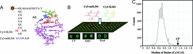
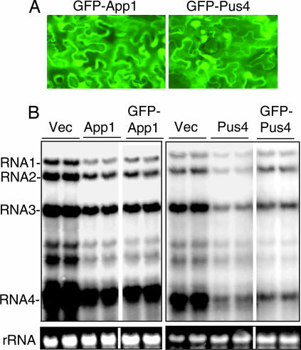
Arrays of >5,000 Saccharomyces cerevisiae proteins were screened to identify proteins that can preferentially bind a small RNA hairpin that contains a clamped adenine motif (CAM). A CAM is required for the replication of Brome Mosaic Virus (BMV), a plant-infecting RNA virus that can replicate in S. cerevisiae. Several hits were selected for further characterization in Nicotiana benthamiana. Pseudouridine Synthase 4 (Pus4) and the Actin Patch Protein 1 (App1) modestly reduced BMV genomic plus-strand RNA accumulation, but dramatically inhibited BMV systemic spread in plants. Pus4 also prevented the encapsidation of a BMV RNA in plants and the reassembly of BMV virions in vitro. These results demonstrate the feasibility of using proteome arrays to identify specific RNA-binding proteins for antiviral activities. Furthermore, the effects of Pus4 suggest that the CAM-containing RNA motif provides a regulatory link between RNA replication and encapsidation.


Conflict of interest statement

The authors declare no conflict of interest.

Figures

Fig. 1.
Fig. 1.
Screening the proteome arrays with an RNA containing a CAM. (A) The sequence of the RNA SLD4 containing the CAM motif is shown along with a structure of the CAM (12). The clamped adenine is residue eight in SLD4 and a substitution to a guanine that abolishes CAM formation in RNA mSLD4 is shown below SLD4. Other nucleotides in mSLD4 are identical to those in SLD4. (B) A scheme illustrating the probing of the protein array. An equal mixture of SLD4 with a 5′ Cy3, and mSLD4 with a 5′ Cy5 was used as the probe. Images of the protein spots of two of the positive hits, Pus4 and App1, and the internal positive control, the BMV CP, are shown below the array. (C) The distribution of the results (the median of Cy3/Cy5 ratios) from the array screen. The signal of BMV CP protein is identified within the graph.
Fig. 2.
Fig. 2.
Characterization of the candidate proteins identified in the protein array experiments. (A) Confirmation of SLD4-protein binding by the 12 candidate proteins using a cross-linking assay. Each cross-linking reaction was performed with 20 μg/ml of each purified candidate protein and 1 nM of SLD4 radiolabled with 32P at its 5′ end. (B) Expression of the S. cerevisiae TyA Gag protein did not significantly affect BMV plus-strand RNA accumulation. The identities of the BMV RNAs are noted to the right of the image of the Northern blot analysis. The images of gel slices below the Northern blots show a cellular rRNA stained with ethidium bromide that serves as loading controls for the RNAs. (C) Effects of expressing the empty vector Qri1, Pus4, or App1 on BMV RNA accumulation. The images are of the Northern blots detecting minus- and plus-strand BMV RNAs, as identified by the symbols to the left of the images. The blots were probed first for minus-strand RNAs to generate the top images, then stripped by boiling in a low salt buffer at 95°C and then probed for plus-strand RNA accumulation. The agarose gel strips contain a cellular rRNA that serves as a loading control. Quantification of RNA accumulations were performed with a phosphorimager.
Fig. 3.
Fig. 3.
Expression of Pus4 and App1 in plants. (A) Fluorescent microscopic images of plant cells expressing GFP-App1 and GFP-Pus4 chimeric proteins. GFP signals were visualized and photographed using a Zeiss Axioplan 2 microscope using GFP optimised FITC filter (at 543 nm excitation and 505- to 530-nm emission). (B) Images of autoradiograms demonstrating that GFP-App1 and GFP-Pus4 are capable of inhibiting BMV plus-strand RNA accumulation. The layout of the gel images is identical to those in Fig. 2B. Two independently derived samples were tested for each manipulation and all of the samples were originally from one blot.
Fig. 4.
Fig. 4.
App1 and Pus4 specifically affect BMV RNA infection in plants. (A) A schematic representation of a N. benthamiana plant used for Agro inoculation and examination of virion formation. (B) App1 and Pus4 inoculated at 0.2 OD595 can decrease spread of BMV in plants. The N. benthamiana leaves from which the samples were prepared are noted at the bottom of the gel images. The larger gel images are of Coomassie blue-stained SDS/PAGE. The samples were plant lysates that had the cell debris removed by a 10,000 × g centrifugation. The bands corresponding to the CP and Rubisco are identified to the right of the gel images. Below the larger gel images are images of Coomassie blue-stained CP from virions that were purified through a sucrose density cushion. (C) Effects of a 1.0 OD595 inoculum of Agrobacterium expressing empty vector, Pus4, or App1. (D) Replication-independent encapsidation of BMV RNA. RNA R3CP, which contains the CP coding sequence and the 3′ UTR of RNA3, is capable of being encapsidated but incapable of replication. R3CP was infiltrated into N. benthamiana along with the CP and either the empty vector, Pus4, or App1. The Western blot result shows signals for the CP in the plant lysates or sucrose density gradient-enriched virions probed with antisera to the BMV CP. Rubisco from the total lysates is shown below the image of the autoradiogram used to purify the virions.
Fig. 5.
Fig. 5.
Mechanism of inhibition by Pus4. (A) The competitive binding of Pus4 and the BMV CP to SLD4. The purified BMV CP was printed on the glass slides as 12 arrays with quadruplicate per array. For each binding assay, duplicate arrays were incubated with Cy3-SLD4 and competitor proteins. The Cy3-SLD4 was premixed with BSA (0.2 mg/ml), Qri1, App1, or Pus4 at 1:1 or 2:1 (protein to SLD4) molar ratios. The median of fluorescence density (F532) was used as binding signal. Each data point was averaged from eight binding signals (±SEM). (B) The binding of Pus4 to SLD4 versus mSLD4. The purified Pus4 were printed on the glass slides as 12 arrays with quadruplicate per array. Each binding assay was performed in duplicate with Cy3-SLD4 or Cy3-mSLD4 at concentrations of 0.2, 0.5, 2.0, and 10 μM. The median of fluorescence density (F532) was taken as the binding signal. Each data point was averaged from binding signals of eight spots (±SEM). (C) The active site of Pus4 is not required for inhibition of the plus-strand BMV RNA3 and RNA4 accumulation. The Agrobacterium cultures expressing Pus4, mPus4, and App1 were infiltrated at an OD595 of 0.5. (D) Electron micrographs of the uranyl acetate-stained images from BMV virion reassembly reactions performed in the presence of BSA or Pus4 at equal concentration to the CP. (Scale bar, 50 nm.) (Insets) Averaged images of 50 individual particles from the reassembly reactions. Numbers at the bottom of the micrographs are the means and one standard error from four independent micrographs of each reassembly reaction.

Similar articles

Cited by

References

    1. Knipe DM. Fields Virology. Baltimore: Lippincott Williams & Wilkins; 2001.
    1. Samuel CE. Clin Microbiol Rev. 2001;14:778–809. - PMC - PubMed
    1. Harris RS, Liddament MT. Nat Rev Immunol. 2004;4:868–877. - PubMed
    1. Akira S, Uematsu S, Takeuchi O. Cell. 2006;124:783–801. - PubMed
    1. Ahlquist P. Nat Rev Microbiol. 2006;4:371–382. - PMC - PubMed

Publication types

MeSH terms