Comparative Study
. 2007 Feb 27;104(9):3324-9.
doi: 10.1073/pnas.0611576104. Epub 2007 Feb 21.

Serial analysis of chromatin occupancy identifies beta-catenin target genes in colorectal carcinoma cells

Affiliations
Comparative Study

Serial analysis of chromatin occupancy identifies beta-catenin target genes in colorectal carcinoma cells

Gregory S Yochum et al. Proc Natl Acad Sci U S A. .

Abstract

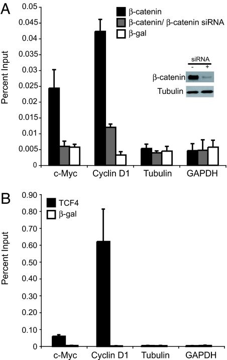
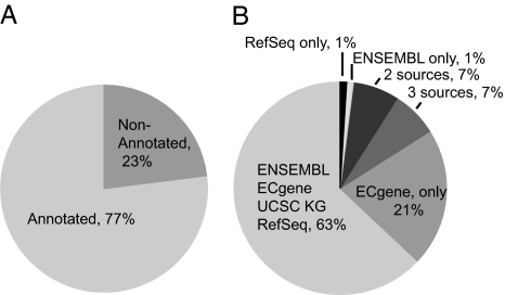
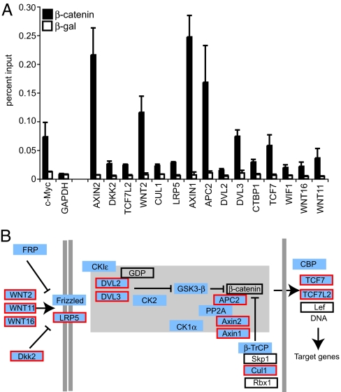
Most instances of colorectal cancer are due to abnormalities in the Wnt signaling pathway, resulting in nuclear accumulation of beta-catenin. beta-Catenin activates transcription of target genes primarily by associating with the T cell factor/lymphoid enhancer-binding factor (TCF/Lef) family of transcription factors. In this report, we use serial analysis of chromatin occupancy (SACO) to identify 412 high-confidence beta-catenin targets in HCT116 colorectal carcinoma cells. Of these targets, 84% contained a consensus TCF motif and were occupied by TCF4 in vivo. Examination of the flanking 5-bp residues in each consensus revealed motif-specific enrichment at neighboring sites. beta-Catenin binding was localized to the 5' promoters, internal regions, and 3' UTRs of protein-coding genes. Furthermore, 15 components of the canonical Wnt pathway were identified as beta-catenin target genes, suggesting that feed-forward and feedback mechanisms exist to modulate the Wnt signal in colon cancer cells.


Conflict of interest statement

The authors declare no conflict of interest.

Figures

Fig. 1.
Fig. 1.
β-Catenin and TCF4 bind c-myc and cyclin D1 promoters in HCT116 colorectal carcinoma cells. (A) Real-time PCR analysis of DNA fragments precipitated in a ChIP assay by using a β-catenin antibody (black bars) or a β-galactosidase antibody (white bars) as a control. Primers designed to the 5′ promoters of c-myc and cyclin D1 were used to detect specific β-catenin binding, and primers designed to the 5′ promoters of tubulin and GAPDH were used to detect nonspecific interactions. As an additional control, cells were incubated with siRNAs to β-catenin for 48 h before ChIP. (Inset) A Western blot demonstrates that β-catenin protein is diminished in siRNA treated cells, whereas tubulin levels remain unchanged. β-Catenin binding to c-myc and cyclin D1 is lowered in siRNA-treated cells (gray bars). (B) The same as in A except that a TCF4 antibody was used in ChIP. In A and B, standard curves were derived from PCRs with serially diluted input chromatin DNA as the template. Data are represented as percent input. Error bars represent SEM.
Fig. 2.
Fig. 2.
Validation of β-catenin-binding sites identified in the SACO library. (A) Real-time PCR quantitation of DNA fragments precipitated in a ChIP assay with a β-catenin antibody (filled bars) or a β-galactosidase antibody as a control (open bars). Genomic targets were chosen at random from 412 loci containing three or more clustered β-catenin GSTs. Primers were designed to a 500-bp region surrounding the mean GST by using primer3 software (Massachusetts Institute of Technology). The mean GST is the midpoint of GST anchors that define the cluster. A locus on the x axis labeled with a known gene symbol indicates that the position of mean GST is within or near 2.5 kb of that gene. All other loci are indicated by the chromosomal position of the mean GST cluster. Data are represented as percent input. Regions interrogated as negative controls included tubulin, GAPDH, the cyclin D1 3′ UTR, and six 5-kb regions of the genome chosen at random, which lacked a consensus TCF motif (CTTTG A/T A/T). (B) The same as in A except that a TCF4 antibody was used to precipitate the putative targets in a ChIP assay. Two independent clusters in the villin 2 gene (VIL2) were confirmed. Error bars represent SEM.
Fig. 3.
Fig. 3.
Distribution of high-confidence β-catenin targets relative to annotation features. (A) Mapping of β-catenin targets containing three or more GSTs within 2.5 kb of annotation features defined in ECgene, Ensembl, RefSeq, and University of California, Santa Cruz known gene databases. Of the targets, 77% align to features in these databases. (B) Assignment of clusters within annotation features listed in A.
Fig. 4.
Fig. 4.
β-Catenin binds targets in the canonical Wnt signaling pathway. (A) Real-time PCR quantitation of fragments precipitated in a ChIP assay using a β-catenin antibody (filled bars) or a β-galactosidase antibody (open bars) as a control. Targets that have significant binding are indicated on the x axis. The amount of DNA precipitated is indicated on the y axis and is relative to levels obtained with input chromatin serially diluted to generate standard curves. The c-myc promoter is a positive control, and GAPDH is a negative control for the ChIP. Error bars represent SEM. (B) Overlay of putative β-catenin SACO targets on the Wnt pathway. The Wnt signaling diagram is a modified version from the Kyoto Encyclopedia of Genes and Genomes (KEGG) available at the Cancer Genome Anatomy Project (CGAP) pathways web page. Targets identified in the SACO library are blue. Targets in blue with a red border were confirmed by ChIP in A. Proteins in an open box were not identified as targets in the SACO library. Proteins in the β-catenin destruction complex are represented within the gray-shaded region.

References

    1. Clevers H. Cell. 2006;127:469–480. - PubMed
    1. Nagase H. Hum Mutat. 1993;2:425–434. - PubMed
    1. Morin PJ, Sparks AB, Korinek V, Barker N, Clevers H, Vogelstein B, Kinzler KW. Science. 1997;275:1787–1790. - PubMed
    1. Ilyas M, Tomlinson IP, Rowan A, Pignatelli M, Bodmer WF. Proc Natl Acad Sci USA. 1997;94:10330–10334. - PMC - PubMed
    1. Nusse R, Varmus HE. Cell. 1982;31:99–109. - PubMed

Publication types