Comparative Study
. 2007 Feb 27;104(9):3336-41.
doi: 10.1073/pnas.0611405104. Epub 2007 Feb 20.

A common variant in combination with a nonsense mutation in a member of the thioredoxin family causes primary ciliary dyskinesia

Affiliations
Comparative Study

A common variant in combination with a nonsense mutation in a member of the thioredoxin family causes primary ciliary dyskinesia

Bénédicte Duriez et al. Proc Natl Acad Sci U S A. .

Erratum in

  • Proc Natl Acad Sci U S A. 2007 Apr 10;104(15):6490

Abstract

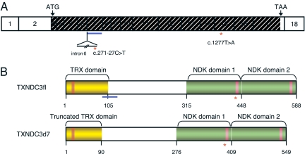
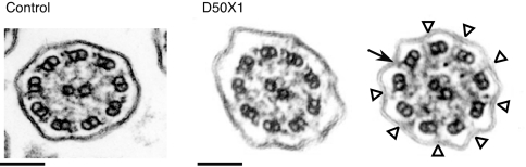
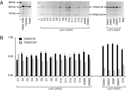
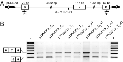
Thioredoxins belong to a large family of enzymatic proteins that function as general protein disulfide reductases, therefore participating in several cellular processes via redox-mediated reactions. So far, none of the 18 members of this family has been involved in human pathology. Here we identified TXNDC3, which encodes a thioredoxin-nucleoside diphosphate kinase, as a gene implicated in primary ciliary dyskinesia (PCD), a genetic condition characterized by chronic respiratory tract infections, left-right asymmetry randomization, and male infertility. We show that the disease, which segregates as a recessive trait, results from the unusual combination of the following two transallelic defects: a nonsense mutation and a common intronic variant found in 1% of control chromosomes. This variant affects the ratio of two physiological TXNDC3 transcripts: the full-length isoform and a novel isoform, TXNDC3d7, carrying an in-frame deletion of exon 7. In vivo and in vitro expression data unveiled the physiological importance of TXNDC3d7 (whose expression was reduced in the patient) and the corresponding protein that was shown to bind microtubules. PCD is known to result from defects of the axoneme, an organelle common to respiratory cilia, embryonic nodal cilia, and sperm flagella, containing dynein arms, with, to date, the implication of genes encoding dynein proteins. Our findings, which identify a another class of molecules involved in PCD, disclose the key role of TXNDC3 in ciliary function; they also point to an unusual mechanism underlying a Mendelian disorder, which is an SNP-induced modification of the ratio of two physiological isoforms generated by alternative splicing.


Conflict of interest statement

The authors declare no conflict of interest.

Figures

Fig. 1.
Fig. 1.
The human TXNDC3 gene and related products. (A) TXNDC3 cDNA structure showing the location of the exons drawn to scale. The translation start and stop codons are labeled with ATG and TAA, respectively. The translated region is hashed. Exon 7 is underlined in blue, and intron 6 is shown as a thin line below exons 6 and 7. The red asterisks mark the locations of the c.271–27C>T and c.1277T>A nucleotide variations, located in intron 6 and exon 15, respectively. (B) Structure of the TXNDC3 isoforms. The TXNDC3fl isoform (Upper), and the TXNDC3d7 isoform (Lower). The thioredoxin (TRX) domain and the two NDK domains are shown in yellow and green, respectively. Within the TRX domain, the active site (GCPC) is shown by an orange box, and, within the NDK domains, the putative NDP kinase active sites are shown by pink boxes. The location of the region encoded by exon 7 is underlined in blue. The location of the p.Leu426X mutation is shown by a red asterisk.
Fig. 2.
Fig. 2.
Electron micrograph of cross-sections of respiratory cilia. (Left) Normal ciliary ultrastructure. (Right) Two cross-sections of respiratory cilia from patient D50X1 showing a heterogeneous ultrastructure: whereas some cilia appear normal (Left), others (66%) display outer dynein arm defects (Right). The black arrow indicates a normal outer dynein arm, and white arrowheads indicate shorter or missing outer dynein arms. (Scale bars: 100 nm.)
Fig. 3.
Fig. 3.
In vivo expression of TXNDC3fl and TXNDC3d7 isoforms. (A) Expression of the TXNDC3fl and TXNDC3d7 isoforms by means of conventional RT-PCR using primers F1 and R1 located in exons 6 and 8, respectively. RT-PCR amplifications were performed on RNAs extracted from human testis, trachea, and nasal cells, and from lymphoblastoid cell lines established for all members of family D50 (D50F, D50M, D50X1, D50S1, and D50S2), for patient D65X1 with a c.271–27C/T genotype, and for 15 controls, C1 to C15, with C1 to C14 having a c.271–27C/C genotype and C15 having a c.271–27C/T genotype. One representative experiment of three independent experiments is shown. L, 1-kb+ ladder; Neg, reaction without RNA. (B) Relative expression of TXNDC3fl and TXNDC3d7 transcripts generated by conventional PCR. The amounts of TXNDC3fl and TXNDC3d7 transcripts, calculated by the GeneTools program, are represented with black and gray bars, respectively. The sum of the two isoforms was arbitrarily set to 1. (Left) The results for individuals who carry the c.271–27C/C genotype, i.e., the patient's mother (D50M) and brother (D50S2) and controls (C1 to C14). (Right) The results for the five individuals bearing the c.271–27C>T variant, i.e., patient D50X1, her father (D50F), her brother (D50S1), patient D65X1, and control C15. Results are represented as means ± SEM of three independent experiments.
Fig. 4.
Fig. 4.
Impact of the c.271–27C>T variant on splicing of TXNDC3 transcripts. (A) Schematic representation of the different TXNDC3 constructs used in this experiment. Exons and introns are represented by open boxes and thin lines, respectively. The location of the c.271–27C/T SNP is indicated by a black arrowhead. The primers used in the RT-PCR experiments (F1 and R1) are represented by horizontal arrows. (B) RT-PCR amplification of TXNDC3 transcripts from HeLa cells transfected with minigenes carrying either a cytosine in intron 6 (i.e., pTXNDC3_C1, pTXNDC3_C2, pTXNDC3_T1>C, and pTXNDC3_T2>C) or a thymine at that position (i.e., pTXNDC3_T1, pTXNDC3_T2, pTXNDC3_C1>T, and pTXNDC3_C2>T) (see Materials and Methods). L, 1-kb+ ladder.

Similar articles

Cited by

References

    1. Afzelius BA. Science. 1976;193:317–319. - PubMed
    1. Kartagener M. Beitr Klin Tuberk. 1933;83:489–501.
    1. Fliegauf M, Omran H. Trends Genet. 2006;22:241–245. - PubMed
    1. Badano JL, Mitsuma N, Beales PL, Katsanis N. Annu Rev Genomics Hum Genet. 2006;7:125–148. - PubMed
    1. Pazour GJ, Agrin N, Walker BL, Witman GB. J Med Genet. 2006;43:62–73. - PMC - PubMed

Publication types