Comparative Study
. 2007 Feb 27;104(9):3366-71.
doi: 10.1073/pnas.0611128104. Epub 2007 Feb 21.

Host shutoff during productive Epstein-Barr virus infection is mediated by BGLF5 and may contribute to immune evasion

Affiliations
Comparative Study

Host shutoff during productive Epstein-Barr virus infection is mediated by BGLF5 and may contribute to immune evasion

Martin Rowe et al. Proc Natl Acad Sci U S A. .

Abstract

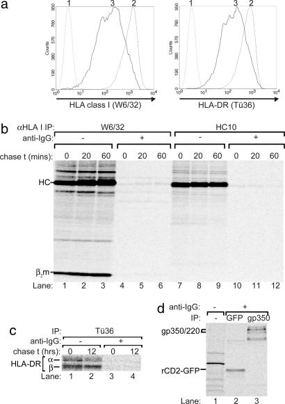
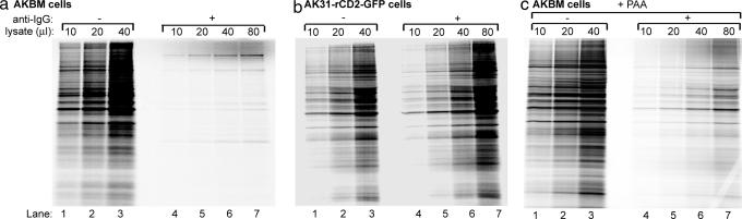
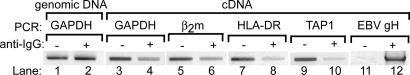
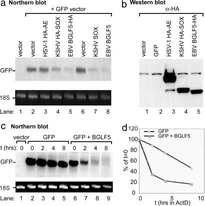
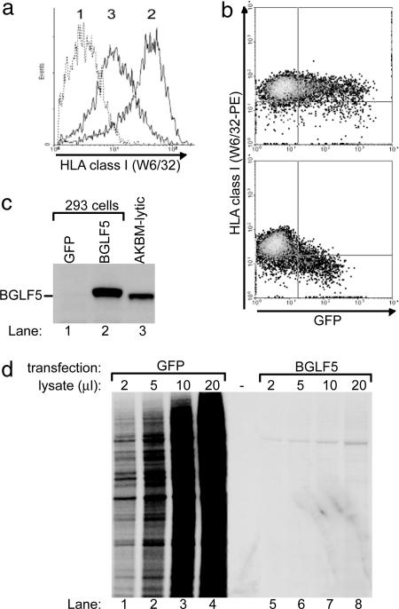
Relatively little is known about immune evasion during the productive phase of infection by the gamma(1)-herpesvirus Epstein-Barr virus (EBV). The use of a unique system to isolate cells in lytic cycle allowed us to identify a host shutoff function operating in productively EBV-infected B cells. This impairment of protein synthesis results from mRNA degradation induced upon expression of the early lytic-cycle gene product BGLF5. Recently, a gamma(2)-herpesvirus, Kaposi sarcoma herpesvirus, has also been shown to encode a host shutoff function, indicating that host shutoff appears to be a general feature of gamma-herpesviruses. One of the consequences of host shutoff is a block in the synthesis of HLA class I and II molecules, reflected by reduced levels of these antigen-presenting complexes at the surface of cells in EBV lytic cycle. This effect could lead to escape from T cell recognition and elimination of EBV-producing cells, thereby allowing generation of viral progeny in the face of memory T cell responses.


Conflict of interest statement

The authors declare no conflict of interest.

Figures

Fig. 1.
Fig. 1.
HLA class I expression is blocked during productive EBV infection. (a) Flow cytometry analysis of latently infected (rat CD2-GFP reporter) and lytically infected (rat CD2-GFP reporter+) AKBM cells at 20 h after induction with anti-IgG. Histograms are depicted for latently (2) and lytically (3) EBV-infected cells showing surface staining for HLA class I (mAb W6/32) and HLA class II (mAb Tü36); as a negative control, no primary antibody was added (1). (b) Latently (−anti-IgG) and lytically (+anti-IgG, sorted 20 h after induction) EBV-infected AKBM cells were metabolically labeled for 1 h with [35S]Met and chased for the times indicated. HLA class I molecules were recovered from Nonidet P-40 cell lysates with conformation-dependent mAb W6/32 (lanes 1–6) or, in the presence of SDS, conformation-independent mAb HC10 (lanes 7–12). Samples were analyzed by SDS/PAGE. (c) In another experiment, AKBM cells were labeled for 6 h with [35S]Cys. HLA class II was immunoprecipitated from Nonidet P-40-cell lysates using mAb Tü36. (d) From the lysates in b, proteins induced in AKBM cells upon viral reactivation were precipitated with Abs specific for GFP (for reporter rat CD2-GFP) and gp350 (mAb 72A1).
Fig. 2.
Fig. 2.
EBV lytic cyle protein(s) expressed at early times of infection induce host shutoff. (a and b) EBV+ AKBM cells (a) and control EBV AK31-ratCD2-GFP cells (b) were cultured with anti-IgG antibody to induce EBV reactivation. At 20 h after induction, AKBM cells in the lytic cycle were positively selected for the expression of the inducible rat CD2-GFP reporter protein; the control AK31 cells constitutively expressing the rat CD2-GFP reporter were also sorted and served as controls. All cell populations were pulse-labeled for 60 (a, lanes 1–7) or 30 min (b, lanes 1–7) with [35S]Met before Nonidet P-40-cell lysis. SDS/PAGE analysis is shown for increasing amounts of total protein extracts denatured in sample buffer. (c) AKBM cells were either left uninduced (−anti-IgG) or were induced overnight (+anti-IgG; sorted for reporter+ cells) in the absence or presence of 300 μg/ml PAA added at the time of induction. Cells were assayed for total cellular protein synthesis by [35S]Met labeling, followed by SDS/PAGE, of titrations of total protein extracts.
Fig. 3.
Fig. 3.
Reduced levels of cellular mRNAs in EBV-producing B cells. Genomic DNA (lanes 1 and 2) and cDNA (lanes 3–12) was prepared from latently (−anti-IgG) and productively (+anti-IgG and sorted) EBV-infected AKBM cells; cDNA was obtained after reverse transcription of total cytoplasmic RNA using random primers. PCR products representing the indicated genes (generated using specific primers) were resolved by agarose gel electrophoresis and visualized by ethidium bromide staining.
Fig. 4.
Fig. 4.
EBV BGLF5 promotes enhanced GFP mRNA turnover. (a) 293T cells were transfected with either empty vector or cotransfected with plasmids expressing GFP and HA-tagged HSV-1 AE, KSHV SOX, or EBV BGLF5 at a 1:4 ratio. Total RNA was extracted from half of the cells 24 h after transfection, 10 μg of RNA was resolved by agarose-formaldehyde electrophoresis, transferred to a nylon membrane, and Northern-blotted with a [32P]-labeled GFP DNA probe. In all Northern blots, the ribosomal 18S RNA served as a loading control. (b) Protein lysates from the other half of the cells were immunoblotted with an HA-specific antibody to detect expression of the HA-tagged viral proteins. (c) 293T cells were transfected with either empty vector or cotransfected with plasmids expressing GFP and EBV BGLF5 at a 1:3 ratio. Forty-eight hours after transfection, cells were treated with actinomycin D (2 μg/ml) for the indicated amount of time to halt transcription. Total RNA was harvested from each sample and Northern-blotted with a [32P]-labeled GFP DNA probe. (d) The half-life of the mRNAs was determined by phosphorimager-based quantification of the blot depicted in c. Values for each band represent the percentage of the respective t = 0 time-point intensity.
Fig. 5.
Fig. 5.
HLA class I down-regulation induced by EBV BGLF5. 293 cells were transfected with pcDNA3-BGLF5-IRES-nlsGFP or an empty control vector. (a and b) At 48 h after transfection, HLA class I expression at the surface of transfected cells was visualized by flow cytometry. (c) GFP+ cells were enriched at 24 h after transfection by using a FACS cell sorter. Protein lysates from the transfected and sorted cells were immunoblotted with a BGLF5-specific polyclonal antiserum (rabbit 120). (d) At 48 h, cells were assayed for total protein synthesis by [35S]Met labeling, followed by SDS/PAGE of titrations of total protein extracts.

Similar articles

Cited by

References

    1. Rickinson AB, Kieff E. In: Fields Virology. Knipe DM, Howley PM, editors. Philadelphia: Lippincott Williams & Wilkins; 2001. pp. 2575–2627.
    1. Amyes E, Hatton C, Montamat-Sicotte D, Gudgeon N, Rickinson AB, McMichael AJ, Callan MF. J Exp Med. 2003;198:903–911. - PMC - PubMed
    1. Hislop AD, Annels NE, Gudgeon NH, Leese AM, Rickinson AB. J Exp Med. 2002;195:893–905. - PMC - PubMed
    1. Yao QY, Rickinson AB, Epstein MA. Int J Cancer. 1985;35:35–42. - PubMed
    1. Roizman B, Pellet PE. In: Fields Virology. Knipe DM, Howley PM, editors. Philadelphia: Lippincott Williams & Wilkins; 2001. pp. 2381–2397.

Publication types

MeSH terms

Substances