Comparative Study
. 2007 Feb 27;104(9):3372-7.
doi: 10.1073/pnas.0608100104. Epub 2007 Feb 20.

NF-kappaB activation by the Toll-IL-1 receptor domain protein MyD88 adapter-like is regulated by caspase-1

Affiliations
Comparative Study

NF-kappaB activation by the Toll-IL-1 receptor domain protein MyD88 adapter-like is regulated by caspase-1

Sinead M Miggin et al. Proc Natl Acad Sci U S A. .

Erratum in

  • Proc Natl Acad Sci U S A. 2013 Oct 22;110(43):17600

Abstract

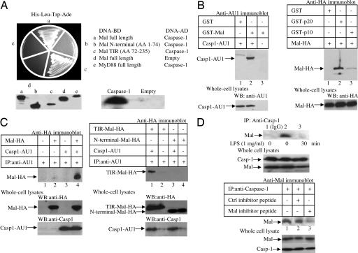
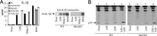
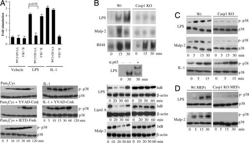
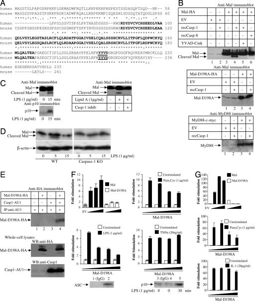
Toll-like receptors (TLRs)-2 and -4 are important proteins in innate immunity, recognizing microbial products and eliciting host defense responses. Both use the adapter proteins MyD88 and MyD88 adapter-like (Mal) to activate signaling pathways. Here we report that Mal but not MyD88 interacts with caspase-1, the enzyme that processes the precursors of the proinflammatory cytokines IL-1beta and IL-18. The interaction was found in a yeast two-hybrid screen and was confirmed by reciprocal GST pull-downs and coimmunoprecipitation of endogenous proteins. We were unable to implicate Mal in regulating caspase-1 activation. However, we found that Mal was cleaved by caspase-1 and that inhibition of caspase-1 activity blocked TLR2- and TLR4-mediated NF-kappaB and p38 MAP kinase activation but not IL-1 or TLR7 signaling, which are Mal independent. These responses, and the induction of TNF, were also attenuated in caspase-1-deficient cells. Finally, unlike wild-type Mal, a mutant Mal, which was not cleaved by caspase-1, was unable to signal and acted as a dominant negative inhibitor of TLR2 and TLR4 signaling. Our study therefore reveals a role for caspase-1 in the regulation of TLR2 and TLR4 signaling pathways via an effect on Mal. This functional interaction reveals an important aspect of the coordination between TLRs and caspase-1 during the innate response to pathogens.


Conflict of interest statement

The authors declare no conflict of interest.

Figures

Fig. 1.
Fig. 1.
Mal interacts with caspase-1. (A) Yeast two-hybrid analysis of Mal, Mal variants, and MyD88 with caspase-1. Immunoblots indicate the presence of respective proteins. (B and C) Interaction of GST-Mal (B Left) or GST-p20 or -p10 (B Right) with lysates from HEK293 cells transfected with either caspase-1-AU1 (B Left) or HA-Mal (B Right) and blotted with relevant antibodies. For immunoprecipitations, HEK293 cells were transfected with plasmids encoding Mal-HA or caspase-1-AU1 (C Left) or encoding TIR-Mal-HA, N-terminal Mal-HA, or caspase-1-AU1 (C Right). (D) THP-1 cells were treated with LPS (top blot) for the indicated times or the Mal inhibitor peptide (20 μM) or a control peptide (20 μM) for 1 h (bottom blot). After immunoprecipitation of the complexes with an IgG control antibody (top blot, lane 1) or an anti-caspase-1 antibody (top blot, lanes 2 and 3, and third blot from the bottom, lanes 1–3), and immunoblotting was performed using an anti-Mal antibody.
Fig. 2.
Fig. 2.
Caspase-1 activation does not require Mal. (A) Wild-type, MyD88-, Mal-, TLR4-, and TLR2-deficient peritoneal macrophages were treated with LPS (100 ng/ml), Malp-2 (0.01 nM), or R848 (0.01 nM) for 16 h followed by ATP (5 mM) for 20 min. IL-1β production was then assessed by ELISA (Left) and Western blot analysis (Right). (B) Wild-type and Mal-deficient bone marrow-derived macrophages were treated with medium or LPS (100 ng/ml) for 16 h followed by ATP (5 mM for 20 min). Lysates were immunoblotted with a caspase-1 anti-p10 antibody. Results shown are representative of three separate experiments.
Fig. 3.
Fig. 3.
Caspase-1 is required for Mal to signal. (A) (Upper) U373 cells were transfected with a 5x NF-κB reporter gene plasmid. Cells were left untreated or pretreated with YVAD-Cmk (100 μM) or IL-1 receptor antagonist (1 μg/ml) for 1 h. Thereafter, cells were untreated or incubated with LPS (1 μg/ml) or IL-1 (100 ng/ml) for 6 h. Shown is the mean relative stimulation of luciferase activity ± SD for a representative experiment from three separate experiments. (Lower) THP-1 cells were left untreated or pretreated with YVAD-Cmk (100 μM) or IETD-Fmk (50 μM) for 1 h followed by treatment with Pam3Cys (1 μg/ml) or IL-1 (1 μg/ml) for 0–120 min. Activation of p38 was analyzed using an anti-phospho-p38-specific antibody. (B) (Top) Time course of NF-κB activation in wild-type and caspase-1-deficient peritoneal macrophages stimulated with LPS (10 ng/ml), Malp-2 (10 nM), and R848 (10 μM) as detected by EMSA. (Middle) Supershift assay was performed by using an anti-p65 antibody for 1 h before analysis by EMSA. Protein:DNA complexes are shown. (Bottom) Wild-type and caspase-1-deficient murine embryonic fibroblast were treated with LPS (100 ng/ml), lipid A (100 ng/ml), or Malp-2 (10 nM) as indicated, followed by immunoblot analysis of the cell lysates with antibodies directed against IκBα or β-actin. (C) Time course of p38 activation in wild-type and caspase-1-deficient peritoneal macrophages stimulated with LPS (100 ng/ml), Malp-2 (10 nM), and IL-1 (1 μg/ml) analyzed by immunoblotting with phospho-p38-specific antibodies. Total p38 levels are also shown. (D) Time course of p38 activation in wild-type and caspase-1-deficient peritoneal murine embryonic fibroblasts stimulated with LPS (100 ng/ml) or Malp-2 analyzed by immunoblotting with phospho-p38-specific antibodies. Total p38 levels are also shown.
Fig. 4.
Fig. 4.
Mal is a substrate for caspase-1. (A) ClustalW alignment of the amino acid sequence of human (top rows) and mouse (lower rows) Mal with the TIR domain in bold and the proposed caspase-1 cleavage site underlined and in bold. (B) for Mal cleavage assay, whole-cell lysates generated from HEK293 cells transfected with a plasmid encoding Mal-HA (Top), Mal-D198A-HA (Middle), or MyD88-cMyc (Bottom) were treated with active recombinant caspase-1 or caspase-8 (Top) in the presence/absence of the caspase-1 inhibitor, YVAD-Cmk. Gels were blotted as indicated. (C) THP1 cells was screened for Mal cleavage following LPS (Left Upper) or Lipid A (± caspase-1 inhibitor; Right Upper) treatment by immunoblotting with an anti-Mal antibody. Cell lysates were also immunoblotted with an antibody against the p10 subunit of caspase-1 (Left Lower). (D) Wild-type and caspase-1 peritoneal macrophages were screened for Mal cleavage after LPS treatment by immunoblotting with an anti-Mal antibody or an anti-β-actin antibody as a loading control. (E) HEK293 cells were transfected with plasmids encoding Mal-D198A-HA or caspase-1-AU1. Immunoprecipitated caspase-1-AU1 was probed for the presence of Mal by immunoblotting. (F) HEK293 cells were transfected with a 5x NF-κB reporter gene plasmid and cotransfected with plasmids encoding empty vector (EV), Mal (1, 40, and 80 ng), or Mal-D198A (1, 40, and 80 ng) (Top Left). HEK293-TLR2 (Top Right), HEK293-TLR4 (Bottom Left), or HEK 293 cells (Bottom Right) were transfected with Mal-D198A (0, 1, 40, and 80 ng) for 24 h. Cells were left untreated or treated with Pam3Cys, LPS, or TNF-α for 6 h. In addition, ASC and the p10 subunit of caspase-1 were immunoprecipitated from untreated (lanes 1–4) or LPS-treated (1 μg/ml, 30 min, lane 5) HEK293-TLR4 cells using an anti-human ASC antibody (lane 2; Genentech) or an anti-p10 antibody (lanes 4 and 5) with an IgG antibody serving as a control (lanes 1 and 3). (G) HEK293 cells were transfected with an IL-8 reporter gene plasmid and cotransfected with plasmids encoding empty vector (EV), Mal (1, 40, and 80 ng), or Mal-D198A (1, 40, or 80 ng) (Top). HEK293-TLR2 (Middle) or HEK293 R1 cells (Bottom) were transfected with Mal-D198A (0, 1, 40, or 80 ng) for 24 h. Cells were left untreated or treated with Pam3Cys or IL-1 for 6 h. For all luciferase assays, mean relative stimulation of luciferase activity ± SD from triplicate determinations for a representative experiment from three separate experiments is shown.

Similar articles

Cited by

References

    1. Miggin SM, O'Neill LA. J Leukoc Biol. 2006;80:220–226. - PubMed
    1. Akira S, Takeda K. Nat Rev Immunol. 2004;4:499–511. - PubMed
    1. Kawai T, Akira S. Cell Death Differ. 2006;13:816–825. - PubMed
    1. Yamamoto M, Sato S, Hemmi H, Sanjo H, Uematsu S, Kaisho T, Hoshino K, Takeuchi O, Kobayashi M, Fujita T, et al. Nature. 2002;420:324–329. - PubMed
    1. Fitzgerald KA, Rowe DC, Barnes BJ, Caffrey DR, Visintin A, Latz E, Monks B, Pitha PM, Golenbock DT. J Exp Med. 2003;198:1043–1055. - PMC - PubMed

Publication types

MeSH terms