Comparative Study
. 2007 Feb 27;104(9):3444-9.
doi: 10.1073/pnas.0608543104. Epub 2007 Feb 20.

Targeted deletion of titin N2B region leads to diastolic dysfunction and cardiac atrophy

Affiliations
Comparative Study

Targeted deletion of titin N2B region leads to diastolic dysfunction and cardiac atrophy

Michael H Radke et al. Proc Natl Acad Sci U S A. .

Abstract

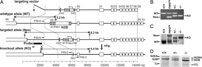
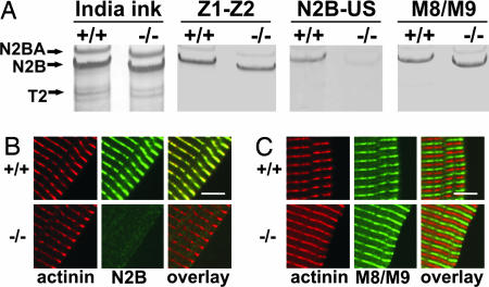
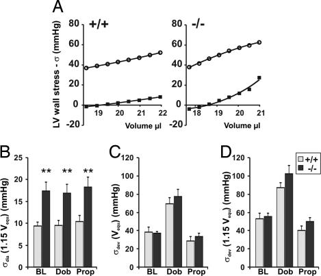
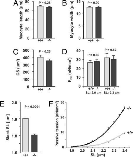
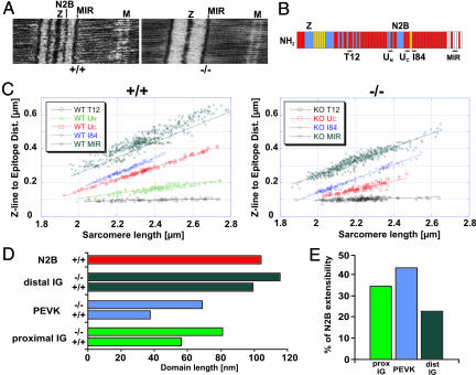
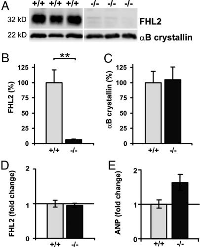
Titin is a giant protein that is in charge of the assembly and passive mechanical properties of the sarcomere. Cardiac titin contains a unique N2B region, which has been proposed to modulate elasticity of the titin filament and to be important for hypertrophy signaling and the ischemic stress response through its binding proteins FHL2 and alphaB-crystallin, respectively. To study the role of the titin N2B region in systole and diastole of the heart, we generated a knockout (KO) mouse deleting only the N2B exon 49 and leaving the remainder of the titin gene intact. The resulting mice survived to adulthood and were fertile. Although KO hearts were small, they produced normal ejection volumes because of an increased ejection fraction. FHL2 protein levels were significantly reduced in the KO mice, a finding consistent with the reduced size of KO hearts. Ultrastructural analysis revealed an increased extension of the remaining spring elements of titin (tandem Ig segments and the PEVK region), which, together with the reduced sarcomere length and increased passive tension derived from skinned cardiomyocyte experiments, translates to diastolic dysfunction as documented by echocardiography. We conclude from our work that the titin N2B region is dispensable for cardiac development and systolic properties but is important to integrate trophic and elastic functions of the heart. The N2B-KO mouse is the first titin-based model of diastolic dysfunction and, considering the high prevalence of diastolic heart failure, it could provide future mechanistic insights into the disease process.


Conflict of interest statement

The authors declare no conflict of interest.

Figures

Fig. 1.
Fig. 1.
Generation of titin N2B-deficient animals. (A) Targeting strategy. The neomycin resistance cassette in the targeting vector replaces the N2B region (exon 49) in the WT allele. The exon encodes three Ig domains and a unique sequence. The Flp recombinase was used to remove the neomycin cassette. The Southern probe is shown as a black box. Restriction sites for Southern blot analysis are EcoRI (E) and BamHI (B). The FRT sites are depicted as gray arrowheads, and primer-binding sites for PCR genotyping are shown as black arrows. (B) PCR genotyping. PCR products derived from the WT allele (+) are 376 bp; targeted allele (neo), 210 bp; KO allele (−), 252 bp. (C) Genomic Southern blot of genomic DNA from the same mice analyzed in B. Digestion of DNA from WT (+/+), heterozygous (neo/+ or +/−), and homozygous (−/−) KO animals with EcoRI and BamHI produces bands of the expected sizes (+, 5.2 kb; neo, 8.2 kb; −, 6.4 kb). (D) Coomassie blue-stained 2% agarose gel with ventricular lysates from +/+, +/−, and −/− animals. N2BA, N2B isoforms, and T2 degradation products are indicated. In the heterozygote, the WT and the mutant proteins (higher mobility) are expressed. The KO expresses only truncated N2B and N2BA titin (tN2BA and tN2B).
Fig. 2.
Fig. 2.
The exon 49-KO affects exclusively the N2B region with proper integration of the truncated protein into the sarcomere. (A) Western blot. India ink staining shows equal loading for WT (+/+) and KO (−/−). Z1-Z2 antibody recognizes the N terminus of titin; the N2B-US antibody, the unique N2B sequence; and the M8/M9 antibody, the C terminus of titin. In KO mice, only the N2B region is deleted, whereas the N- and C-terminal parts of the titin protein are unaffected. (B) Confocal microscopy of double-immunofluorescence-stained cardiomyocytes. Anti-α-actinin (red), labels the Z-disk, and anti-N2B (green) localizes at the I-band close to the Z-disk in WT animals (+/+). In KO mice, the N2B staining is absent. (C) Costaining with anti-α-actinin (red) and the anti-M8/M9 (green) antibody directed against the M-band region of titin shows proper integration of N2B-deficient titin into the sarcomere (alternating actinin/M-band titin staining). (Scale bar, 5 μm.)
Fig. 3.
Fig. 3.
N2B-deficient hearts have increased diastolic wall stress. (A) Examples of σ − V relation in WT (Left) and KO mice (Right). Diastolic stress is shown by squares and developed stress by circles. (B) Mean ± SEM of WT (light gray) and KO (dark gray) mice (six animals each). Results are shown at Veq + 15%, under baseline conditions (BL) and in the presence of either dobutamine (Dob) or propranolol (Prop). Under all conditions, diastolic wall stress is increased in KO mice (P < 0.01). (C and D) Developed wall stress at Veq and Veq + 15%. Developed wall stress is largely unchanged between the genotypes. There is a trend toward higher developed σ in the presence of dobutamine, which did not reach statistical significance (P = 0.08). The corresponding pressures are provided in SI Fig. 9.
Fig. 4.
Fig. 4.
Slack sarcomere length (SL) is reduced, and passive tension is increased in skinned cardiomyocytes of N2B-KO mice. Myocyte length (A), width (B), cross-sectional area (C), and maximal force produced (D; tested at two sarcomere lengths) are unchanged in N2B-KO (−/−) versus WT (+/+) control animals. (E) Slack sarcomere length is significantly reduced in KO versus WT cardiomyocytes (n = 30 each). (F) Passive tension is increased in KO versus WT at sarcomere length >2 μm. Myocytes were derived from age-matched KO (n = 11) and littermate control WT animals (n = 10) at 6 months of age.
Fig. 5.
Fig. 5.
Compensation for loss of the N2B element by the PEVK and tandem Ig regions as determined by immunoelectron microscopy. (A) WT (+/+) and KO (−/−) LVs were colabeled with anti-N2B and anti-MIR antibody. The N2B labeling is only observed in the +/+ and absent in the −/− animal. (B) Antibody-binding sites are shown along the titin I-band region. (C) Measurement of epitope distance from the Z-line at various sarcomere lengths tested on +/+ and −/− muscle strips. (D) Segment extension at sarcomere length 2.3 μm. Tandem Ig and PEVK segments are increased in length in KO mice. (E) Increased extension of tandem Ig and PEVK segments in KO mice as a percentage of extension of I-band titin. Loss of the N2B element results in the largest extension of PEVK segment followed by the proximal tandem Ig and the distal tandem Ig region.
Fig. 6.
Fig. 6.
FHL2 protein level is decreased in the N2B-KO, αB-crystallin is unchanged, and ANP expression is increased. (A) The titin N2B-binding proteins FHL2 and αB-crystallin were quantified by Western blot analysis of WT (+/+) and KO (−/−) LVs (n = 3 each). Although αB-crystallin is unchanged, FHL2 levels are reduced significantly in KOs (−/−). (B and C) Quantification of FHL2 and αB-crystallin normalized to actin levels and (+/+) set to 100%. The difference is significant at P < 0.01 (n = 6 per genotype). (D and E) FHL2 RNA levels as determined by real-time quantitative RT-PCR from left ventricle are unchanged (D), but the hypertrophy marker ANP (E) is up-regulated in KO animals (n = 3 per genotype).

Similar articles

Cited by

References

    1. Granzier HL, Labeit S. Circ Res. 2004;94:284–295. - PubMed
    1. Labeit S, Kolmerer B. Science. 1995;270:293–296. - PubMed
    1. Trombitas K, Freiburg A, Centner T, Labeit S, Granzier H. Biophys J. 1999;77:3189–3196. - PMC - PubMed
    1. Linke WA, Rudy DE, Centner T, Gautel M, Witt C, Labeit S, Gregorio CC. J Cell Biol. 1999;146:631–644. - PMC - PubMed
    1. Matsumoto Y, Hayashi T, Inagaki N, Takahashi M, Hiroi S, Nakamura T, Arimura T, Nakamura K, Ashizawa N, Yasunami M, et al. J Muscle Res Cell Motil. 2005;26:367–374. - PubMed

Publication types

MeSH terms