Comparative Study
. 2007 Feb 27;104(9):3466-71.
doi: 10.1073/pnas.0611271104. Epub 2007 Feb 20.

Identification of IGF2 signaling through phosphoinositide-3-kinase regulatory subunit 3 as a growth-promoting axis in glioblastoma

Affiliations
Comparative Study

Identification of IGF2 signaling through phosphoinositide-3-kinase regulatory subunit 3 as a growth-promoting axis in glioblastoma

Liliana Soroceanu et al. Proc Natl Acad Sci U S A. .

Abstract

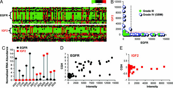
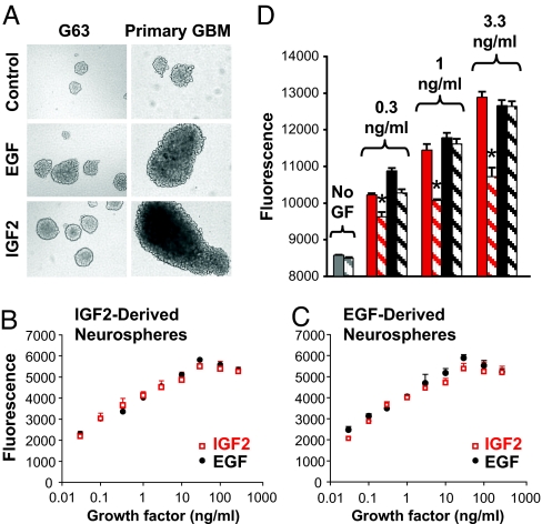
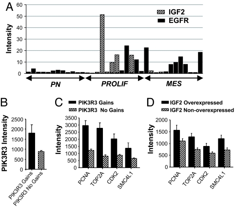
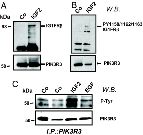
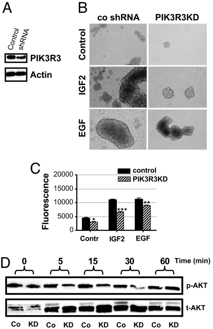
Amplification or overexpression of growth factor receptors is a frequent occurrence in malignant gliomas. Using both expression profiling and in situ hybridization, we identified insulin-like growth factor 2 (IGF2) as a marker for a subset of glioblastomas (GBMs) that lack amplification or overexpression of EGF receptor. Among 165 primary high-grade astrocytomas, 13% of grade IV tumors and 2% of grade III tumors expressed IGF2 mRNA levels >50-fold the sample population median. IGF2-overexpressing tumors frequently displayed PTEN loss, were highly proliferative, exhibited strong staining for phospho-Akt, and belonged to a subclass of GBMs characterized by poor survival. Using a serum-free culture system, we discovered that IGF2 can substitute for EGF to support the growth of GBM-derived neurospheres. The growth-promoting effects of IGF2 were mediated by the insulin-like growth factor receptor 1 and phosphoinositide-3-kinase regulatory subunit 3 (PIK3R3), a regulatory subunit of phosphoinositide 3-kinase that shows genomic gains in some highly proliferative GBM cases. PIK3R3 knockdown inhibited IGF2-induced growth of GBM-derived neurospheres. The current results provide evidence that the IGF2-PIK3R3 signaling axis is involved in promoting the growth of a subclass of highly aggressive human GBMs that lack EGF receptor amplification. Our data underscore the importance of the phosphoinositide 3-kinase/Akt pathway for growth of high-grade gliomas and suggest that multiple molecular alterations that activate this signaling cascade may promote tumorigenesis. Further, these findings highlight the parallels between growth factors or receptors that are overexpressed in GBMs and those that support in vitro growth of tumor-derived stem-like cells.


Conflict of interest statement

The authors declare no conflict of interest.

Figures

Fig. 1.
Fig. 1.
EGFR-OE and IGF2-OE are nonoverlapping across human GBM samples. (A) Heat map displaying microarray data for EGFR (Upper) and IGF2 (Lower) in a set of GBMs. Z score-normalized intensity values are depicted for Affymetrix probes to EGFR or IGF2 as mapped to chromosomes 7p and 11p, respectively. (B) Plot representing the Affymetrix intensity values for EGFR and IGF2 in grade III gliomas (filled symbols) and GBMs (open circles). No overlap occurs between EGFR-OE and IGF2-OE cases. Dashed lines correspond to cut-off values for IGF2-OE (red) and EGFR-OE (black). (C) Normalized mRNA levels (abundance relative to that of Rab14) for EGFR and IGF2 measured by Taqman in 12 selected cases. (D and E) Comparison of log2 CGH ratios vs. expression values for EGFR (D) and IGF2 (E).
Fig. 2.
Fig. 2.
Histological confirmation of nonoverlap between IGF2-OE and EGFR-OE GBMs. (A–D) IGF2 mRNA detection by ISH. (A and C) PhosphorImager scans of tissue microarrays hybridized for IGF2 mRNA using IGF2-antisense (A) or sense strand control probe (C). (B and D) Dark-field microphotographs of ISH of the tissue core indicated by red boxes in A and C. (Scale bar: 1 mm.) (E–J) Tissue sections from an EGFR-positive case (E–G) and an IGF2-positive case (H–J) showing IHC for EGFR, Ki-67, and p-akt. (Scale bar: 100 μm.)
Fig. 3.
Fig. 3.
IGF2 can substitute EGF to support tumor-derived neurosphere growth. (A) (Left) G63 cell line was grown under basal control neurosphere conditions (Top), in the presence of 20 ng/ml EGF (Middle), or with 20 ng/ml IGF2 (Bottom). (Right) Similar results were obtained with neurospheres derived from primary GBM tissue. (B and C) Proliferation assay of cells from neurospheres formed in the presence of IGF2 (B) or EGF (C) shows equivalent growth effects for the two factors. (D) IGF1R blocking antibody (α-IR3, 10 μg/ml) partially inhibits IGF2-induced cell proliferation (∗, P < 0.03) but not the EGF-induced cell growth. Red bars, IGF2; black bars, EGF; solid bars, growth factor alone; striped bars, growth factor plus α-IR3. In B–D representative results of three experiments per cell line are shown. Data are presented as mean ± SD of triplicate samples.
Fig. 4.
Fig. 4.
IGF2 and PIK3R3 are overexpressed in proliferative GBMs. (A) Stacked bar graph shows intensity values for IGF2 and EGFR in 36 samples representing three molecular subtypes of high-grade glioma: proneural (PN), proliferative (PROLIF), and mesenchymal (MES). Values plotted represent intensity values of EGFR or IGF2 for each tumor normalized to the mean intensity of the corresponding gene across all cases. (B and C) Affymetrix intensity values for both expression of PIK3R3 (B) and the proliferative markers PCNA, TOP2A, CDK2, and SMC4L1 (C) are all significantly elevated in cases with PIK3R3 genomic gains compared with those with no gain (n = 6 and 82, respectively; P < 0.001, t test; all comparisons.) (D) Expression of proliferative markers is elevated in IGF2-OE cases (n = 17) compared with IGF2-nonoverexpressing samples (n = 148; P < 0.05, t test; all comparisons).
Fig. 5.
Fig. 5.
IGF2 treatment induces association between phopho-IGF1R and PIK3R3 in glioma cells. Western blot show PIK3R3 immunoprecipitates of G96 cells after IGF2 stimulation. Cells were stimulated with IGF2 (20 ng/ml, 30 min) or EGF (10 ng/ml, 30 min) or were unstimulated (labeled Co). Blots were probed with antibodies as follows: antibody to IGF1R kinase domain (A Upper); antibody to PIK3R3 (A Lower); antibody to phospho-IGF1R (PY1158/Y1162/Y1163) (B Upper); antibody to PIK3R3 (B Lower); antibody to phospho-tyrosine (C Upper); antibody to PIK3R3 (C Lower).
Fig. 6.
Fig. 6.
PIK3R3 KD inhibits IGF2-induced growth of glioma-derived neurospheres. (A) Western blot reveals that stable PIK3R3 KD in G96 cells decreases protein levels compared with control shRNA-treated cells. (B) Representative neurospheres of PIK3R3KD and control cells demonstrate that PIK3R3KD inhibits sphere formation and/or growth induced by either EGF or IGF2 in G96 cells. (C) Viability assays of neurosphere growth show a decrease in the number of PIK3R3KD cells that is most profound in IGF2-stimulated cultures (∗, P < 0.05; ∗∗, P < 0.005; ∗∗∗, P < 0.0005). (D) In the absence of growth factor stimulation, p-akt levels are equivalent in control (Co) and KD cells. Under IGF2 (20 ng/ml) stimulation, p-Akt levels in G96PIK3R3KD cells are decreased compared with G96 control cells.

Similar articles

Cited by

References

    1. Holland EC. Nat Rev Genet. 2001;2:120–129. - PubMed
    1. Galli R, Binda E, Orfanelli U, Cipelletti B, Gritti A, De Vitis S, Fiocco R, Foroni C, Dimeco F, Vescovi A. Cancer Res. 2004;64:7011–7021. - PubMed
    1. Sanai N, Alvarez-Buylla A, Berger MS. N Engl J Med. 2005;353:811–822. - PubMed
    1. Nutt C, Louis DN. Cancer of the Nervous System. 2nd Ed. New York: McGraw–Hill; 2005. pp. 837–847.
    1. Phillips HS, Kharbanda S, Chen R, Forrest WF, Soriano RH, Wu TD, Misra A, Nigro JA, Colman H, Soroceanu L, et al. Cancer Cell. 2006;9:157–163. - PubMed

Publication types