Comparative Study
. 2007 Feb 27;104(9):3478-83.
doi: 10.1073/pnas.0611492104. Epub 2007 Feb 20.

Mice expressing a humanized form of VEGF-A may provide insights into the safety and efficacy of anti-VEGF antibodies

Affiliations
Comparative Study

Mice expressing a humanized form of VEGF-A may provide insights into the safety and efficacy of anti-VEGF antibodies

Hans-Peter Gerber et al. Proc Natl Acad Sci U S A. .

Abstract

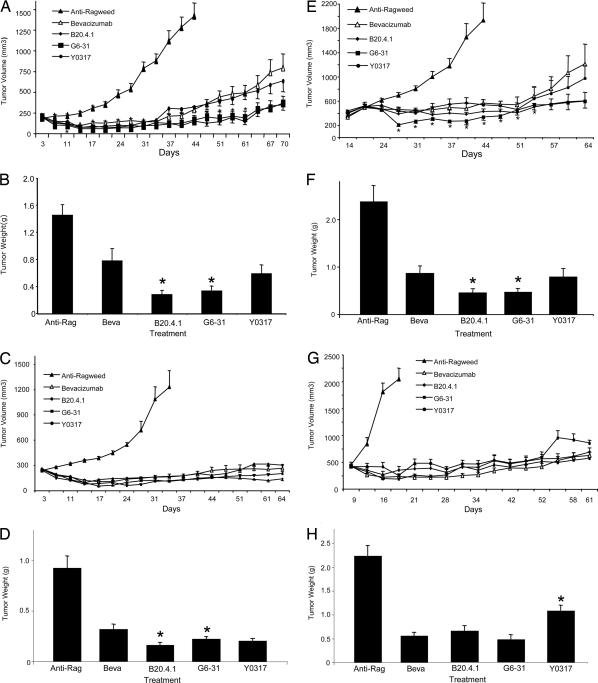
VEGF-A is important in tumor angiogenesis, and a humanized anti-VEGF-A monoclonal antibody (bevacizumab) has been approved by the FDA as a treatment for metastatic colorectal and nonsquamous, non-small-cell lung cancer in combination with chemotherapy. However, contributions of both tumor- and stromal-cell derived VEGF-A to vascularization of human tumors grown in immunodeficient mice hindered direct comparison between the pharmacological effects of anti-VEGF antibodies with different abilities to block host VEGF. Therefore, by gene replacement technology, we engineered mice to express a humanized form of VEGF-A (hum-X VEGF) that is recognized by many anti-VEGF antibodies and has biochemical and biological properties comparable with WT mouse and human VEGF-A. The hum-X VEGF mouse model was then used to compare the activity and safety of a panel of VEGF Mabs with different affinities for VEGF-A. Although in vitro studies clearly showed a correlation between binding affinity and potency at blocking endothelial cell proliferation stimulated by VEGF, in vivo experiments failed to document any consistent correlation between antibody affinity and the ability to inhibit tumor growth and angiogenesis in most animal models. However, higher-affinity antibodies were more likely to result in glomerulosclerosis during long-term treatment.


Conflict of interest statement

Conflict of interest statement: The manuscript describes the use of several anti-VEGF antibodies, including bevacizumab. The authors are employees of Genentech, Inc., the manufacturer of bevacizumab.

Figures

Fig. 1.
Fig. 1.
Ten amino acids mutated from mouse to human to generate the hum-X VEGF variant. Sequence comparison between mouse and human VEGF-A. A total of 19 aa are different between murine VEGF164 and human VEGF165 (shaded gray). Ten amino acids (boxed and gray) located within exons 3, 4, and 5 of mouse VEGF were mutated to human residues by site-directed mutagenesis.
Fig. 2.
Fig. 2.
Efficacy of anti-VEGF-A Mabs in human tumor xenografts implanted s.c. into RAG2 KO; hum-X VEGF KI double-homozygous mice. (A–D) Prevention studies. (E–H) Intervention studies. (A) Growth curves of Calu-6 tumors. Control anti-Ragweed (Anti-Rag), B20–4.1, G6–31, bevacizumab (Beva) or Y0317 Mabs were administered at 5 mg/kg, i.p., twice weekly. (B). Terminal weights of Calu-6 tumors from experiment in A at day 70 of treatment. Control animals were killed at day 44. Tumors from B20-4.1- and G6-31-treated animals had significantly lower weight than the bevacizumab group. (C) Growth curves of human HT29 colorectal carcinoma cells. (D) Terminal weights of HT29 tumors from the experiment in C as determined at day 67. Controls were killed at day 33. (E) Growth curves of Calu-6 tumors in which treatment was initiated after tumor volumes reached ≈400 mm3 (regression study). (F) Terminal tumor weights of Calu-6 tumors in E were determined at day 63. Controls were harvested at day 42. (G) Growth curves of human HM7 colorectal tumor. Similar to E and F, antibody treatment was initiated after tumor volumes reached ≈400 mm3. (H) Terminal weights of HM7 tumors determined on day 61. Controls were killed at day 19. Data shown are Means ± SEM. ∗, Significant difference (P < 0.05) compared with the bevacizumab group.
Fig. 3.
Fig. 3.
Renal changes in homozygous RAG2 KO; hum-X VEGF KI double-homozygous mice treated with anti-VEGF-A antibodies (human Fc framework). Animals treated (5 mg/kg, two times weekly for 54 days) with antibodies having increasing affinity for VEGF-A have increased glomerulosclerosis with expanded mesangial areas and thickened capillary loops (A–E). Anti-murine VEGF staining (F–J); control animals (F) show moderate podocyte-specific staining, without capillary loop or mesangial signal; treatment with antibodies of increasing affinity (G–J) results in progressively increased signal in mesangial areas and capillary loops; capillary loop staining in variably linear (J, Y0317) or more coarsely granular (I, G6–31). VEGF-A signal in juxtamedullary glomeruli is consistently stronger than in peripheral cortical glomeruli (detail not shown). Anti-human Fc (K–O, direct immunofluorescence); anti-VEGF antibodies of increasing affinity accumulate in glomeruli roughly in proportion to their affinity. Complement C3 (P–T, direct immunofluorescence); anti-VEGF antibodies of increasing affinity result in complement C3 deposition in glomeruli, roughly in proportion to their affinity; nonspecific signal is present in Bowman's capsule basement membrane surrounding glomerular tuft.
Fig. 4.
Fig. 4.
Renal ultrastructural changes in homozygous RAG2 KO; hum-X VEGF KI double-homozygous mice treated with anti-VEGF-A antibodies (murine Fc framework). (A and B) Treatment with G6–31 5 mg/kg, weekly for 8 weeks, results in diffusely increased mesangial cellularity, widening of mesangium and capillary loops by amorphous extracellular material (1-μm methacrylate sections; Jones silver stain (A) and toluidine blue (B). (C and D) Treatment with G6–31 10 mg/kg, twice weekly for 8 weeks, results in variably distorted EC, focally enveloping amorphous subendothelial deposits (asterisks indicate presumably immune complexes); EC frequently lack regularly spaced fenestrations. Basement membrane shows focal reduplication (arrows). Podocyte foot processes are variably fused.

References

    1. Ferrara N. Endocr Rev. 2004;25:581–611. - PubMed
    1. Ferrara N, Hillan KJ, Gerber HP, Novotny W. Nat Rev Drug Discov. 2004;3:391–400. - PubMed
    1. Gasparini G, Longo R, Toi M, Ferrara N. Nat Clin Pract Oncol. 2005;2:562–577. - PubMed
    1. Ferrara N, Kerbel RS. Nature. 2005;438:967–974. - PubMed
    1. Jain RK, Duda DG, Clark JW, Loeffler JS. Nat Clin Pract Oncol. 2006;3:24–40. - PubMed

Publication types

MeSH terms

Substances

LinkOut - more resources